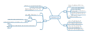
导图社区 幼儿保育VR课件脚本-幼儿生活中常见问题
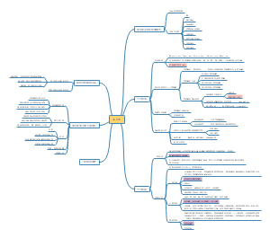
幼儿保育VR课件脚本-幼儿生活中常见问题
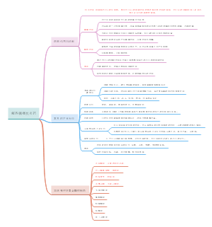
拿出电话(奶奶带电话的姿势即可,减少人物动作。),“喂?宝贝今天在家玩的时候,不小心摔伤了,腿部蹭破一点皮。我已经做了简单的处理了,马上再去医院给孩子看看,你们下班之后也过来吧!我在小区门口的医院。”
编辑于2022-10-25 02:36:53 北京市- 幼儿
- 幼儿保育
- VR课件开发
- 相似推荐
- 大纲
导图社区 幼儿保育VR课件脚本-幼儿生活中常见问题
拿出电话(奶奶带电话的姿势即可,减少人物动作。),“喂?宝贝今天在家玩的时候,不小心摔伤了,腿部蹭破一点皮。我已经做了简单的处理了,马上再去医院给孩子看看,你们下班之后也过来吧!我在小区门口的医院。”
编辑于2022-10-25 02:36:53 北京市