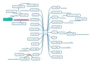
导图社区 Host cell I and II
Host cell I and II
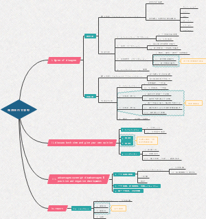
Host cell I and II思维导图:包含Cellular homeostasis,Proteasome - antigen presentation or protein recycling ,These quality control measures generally lead to the degradation of mis-folded or foreign proteins等等
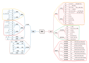
编辑于2022-05-10 13:44:24- B cell Immunodeficiency
这是一篇关于B cell Immunodeficiency的思维导图,其内容包括X—linked agammaglobulinemia(XLA),Hyper Igm syndrome等四个方面的内容
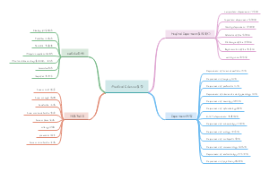
- NK Cells
这是一篇关于Natrual Killer Cells免疫学 墨尔本大学的思维导图,对于NK Cells感兴趣的小伙伴可以收藏起来。
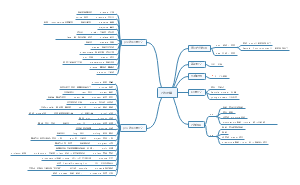
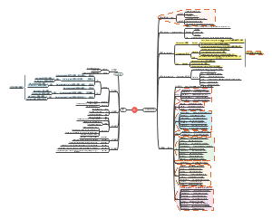
- Bacteria Pathogenesis
Bacteria Pathogenesis思维导图:包含Normal Microbiota,Difference between Pathogens &Normal microbiota,Enter the body,Colonisation of the Host,Invasion of the host cell等等
Host cell I and II
社区模板帮助中心,点此进入>>
- B cell Immunodeficiency
这是一篇关于B cell Immunodeficiency的思维导图,其内容包括X—linked agammaglobulinemia(XLA),Hyper Igm syndrome等四个方面的内容
- NK Cells
这是一篇关于Natrual Killer Cells免疫学 墨尔本大学的思维导图,对于NK Cells感兴趣的小伙伴可以收藏起来。
- Bacteria Pathogenesis
Bacteria Pathogenesis思维导图:包含Normal Microbiota,Difference between Pathogens &Normal microbiota,Enter the body,Colonisation of the Host,Invasion of the host cell等等
- 相似推荐
- 大纲