导图社区 Cell signaling 2.0
- 41
- 1
- 0
- 举报
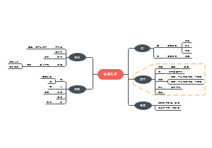
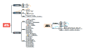
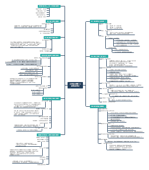
Cell signaling 2.0
Cell signaling 2.0:Primary messenger、The Receptors、Secondary messengers、Modulator of the secondary messengers……
编辑于2022-06-09 23:04:17- cell
- Anchoring
- Modulato…
- Downstre…
- Modulator
- 相似推荐
- 大纲

Cell signaling
Primary messenger
Signals in broad sense
Antigens
Cell surface glycoproteins+oligosaccharides
Developmental signals
Extracellular Matrix components
Growth factors
Hormones
Light
Mechanical touch
Neurotransmitters
Nutrients
Oderants
Pheromones
Tastants
Chemistry
Proteins
glucagon
4.5 kDa
Growth factors
5-50 kDa
EGF
EGF has MW of 6kDa, which is stabalized by its internal disulfide bonds
It is derived from the cleavage of a large transmembrane protein
insulin
Gas
NO
diffuse rapidly, but the effects is short because its turn over is rapid
Small organic molecules
lipids
estrogen
aminoacids
Adrenaline
others
Dopamine
Functions
Neurotransmitters
NO
Dopamine
Glutamate
Serotonin
Hormones
Epinephrine
also called adrenaline
Released from adrenal gland
regulates energy metabolisms in
muscle
liver
adipose tissue
serves as neurotransmitter in
adrenergic neurons
Synthetic analogs
Isoproterenol
agonist with slightly higher affinity
propranolol
antagonist with extremely high affinity
Steroid
Pheremones
Paracrine signals
cytokines
Interferon(IFN)
all cells can synthesis when viral infection
IFN-α
antiviral
IFN-β
antiviral
They induce neighboring cell death
by induce neighboring cells RNA degredation and blocks protein production
Only produced by T lymphpcytes and NK cells
IFN-γ
activates macrophage
immonological defense against infection and cancer
They also connect lymphocytes' prolyferation
Interleukine(IL)
complex signaling functions
trigure favor, inflammation, T cells differenciation
IL-4
activates B cell clonal expansion
released by Th1 MHCII recognation
IL-12
enhance Macrophages phagocytosis
released by Th0 MHCII recognation
Chemokines
a family of small cytokines
they are common in inducing chemotaxis of cells
ex. Cb3 in complement system, attract neutrophiles and Macrophages, also C5a attracts neutrophiles
usually attracts phagocytes。。。
Complexity
The Same ligand can has different receptors and even the same receptor is stimulated, it can generates different responses!
Actetylcholine let heart muscle to relax
The same receptor in salivary gland results in slivar secrection
The different receptor in skeletal muscle cells make it contraction
The Receptors
Synthetic ligands
agonist
structural analogs that bind to a receptor, change it conformation and activates the cell's biochemical response
complete agonist
double the effects of the partial agonist
partial agonist
mimic the effects of its natural ligand
reverse agonist
reduces the response to smaller than the base level
antagonist
structural analogs that bind the receptor without triggering the normal effect(no or different in conformational changes) and thereby block the effects of agonists
Affinity
the synthetic agonist and antagonist usually have stronger affinity than the natural ligand
The aminoacids responsed for the interactions with agonist and antagonist are usually shared but not indentical
GPCR(G-protein coupled receptor)
example
adrenergic receptor
α-adrenergic receptor
β-adrenergic receptor
used as the standard GPCR model
Increase breakdown of glycogen and fat
muscle
liver
adipose tissue
Rhodopsin
Variations
350 variations
detect endogenous ligands
hormones
growth factors
500 variations
detect olfactory and gustatory receptors
Functions
diseases
allergies
depression
blindness
diabetes
cardiovescular defects
More than harf of all the drugs are targeting GPCRs!
β-blockers
hypertension
cardiac arrhythmia
glaucoma
anxiety
migraine headache
More than 150 are still functionally unknown!
3 essential components(in β adrenergic receptor)
A receptor with 7 transmembrane helical domain
hydrophobic
with each 20-28 amino acids
The binding site of epinephrine is deep within the plasma membrane
promotes the conformational change in the receptor's intracellular domain, and catalyzes
heterotrimeric G protein (guanosine nucleotide-binding protein)
Gs
heterotrimeric
αβγ
activate when GTP binds to it nucleotide binding site
β and γ subunits dissociate from α subunit as a βγ dimmer
Gsα with its bound GTP move in the plane of the membrane from the receptor to stimulate a near by Adenyl cyclase
Gsa is held to membrane by a covalently attached palmitoyl group
inactivated when GDP binds to it
effector enzyme in the plasma membrane
The down streams of it
The G protein independent signaling is done by arrestin, which activates ERK 1,2(MAPK) and activates the MAP kinase pathway.
Enzyme-linked receptors
Common
All single transmembrane domain
2 common organizations
The Phosphorylated tyrosines on the receptors are always bind to SH2 domains of other proteins
SH2 domain can recognizes phosphotyrosine
RTK(Receptor tyrosin kinase)
Insulin receptors
disulphide bonded tetramer
2 Insulin}extracellular domain---->cross phosphorylation
can be viewed as already dimmerized
EGF (epidermal growth factor) receptor
Organization
only exists in multicellular organisms
EGF can stimulates the growth of epidermal cells
EGF recsptor is a single polypeptide chain containing 1186 aminoacids
Dimerization
EGF}extracellular domain---->receptor dimerization----> cross phosphorylation
dimerized through dimerization arm
cross phosphorylation results in a correct conformational change that brings the tyrosine into the kinase active site
Down stream
Grb-2 (growth factor-receptor bound protein-2)
contains a SH2 domain flanked by 2 SH3 domains
SH3 binds the C-terminal prolin rich sequences on Sos
It central domain has guanosine nucleotide exchange factor!
Its C-terminal has proline rich segments
Sos recruits and activates the membrane anchored G-protein: Ras
Ras
Will finally activates ser/Thr kinases raf through direct interactive, and it activates certain transcription factors through MAPK kinases cascade
MAPKK is also known as MEK(MAPK/ERK kinase)
It is activated by phosphorylation of specific Tyr+Thr residues
It shows dule specificities to phosphorylates 2 kinds of residues
MAPK also have other names like
ERKs (extracellular-signal regulated protein kinases)
MAP kinases/mitogen-activated protein
MAPK will migrates to nucleus and activates transcription factors: Jun, Fos, and Myc.
FGF (fibroblast growth factor) receptor
Functions of FGF
Angiogenesis
Wood healing
Embryonic development
Domain organization
3 Ig like domains, and the D2 and D3 are ligand binding domains, there is an acid box between D1 and D2 & D3
Down stream signaling
How it is activated?
heparin is tetrasaccharide and shows some activity
hexasaccharide is the minimum amount for crystallography
decasaccharide(10) is the optimum amount for activation
All the extracellular matrix coactivators for FGFR
The order they bind
Anosmin+HS
The anosmin must first binds to HS, when it first binds to the receptor(it can), it will inhibits the HS from further binding.
Proteins
E-selectin (an integral membrane protein, involved in cell adhesion, binds FGFs, effect unknown)
Cadherins (Ca2+-dependent adhesion, bind FGFR and signal through these)
Neuropilin-1 (acts co-receptor for FGF, increases cell growth responses)
Anosmin-1 (a secreted extracellular matrix-associated glycoprotein responsible for normal development, altered cell growth responses)
Sugars
Heparan sulphate proteoglycans-HSPG (specialised glycoprotein family)
HS attached on HSPG has same structure with the "heparin", with repeated units of sulfated disaccharide
More specificaly, the disaccharide is variably-sulfated uronic acid (D-glucuronic or l-iduronic acid) and glucosamine (N-acetylated or N-sulfated) monosaccharides
specific sugar sequences sulfation/acetylation/epimerisation patterns
confer selective protein-binding properties.
HS binding FGF>dimerisation of FGF>together form ternary complex with FGFR
3 detailed models proposed
1. Conformational Change
HS{FGF->conformational change of FGF->high affinity and activation of FGFR
HS binding can increase FGF's thermal stability
2. Growth hormone
1*FGF{1*FGFR, the HS then recruits another FGFR, the FGF is bivalent, and HS function as a glue.
site directed mutagenesis suggests the FGF is bivalent
3. Ligand induced dimerization
2FGF,2FGFR
The HS could be 1/2
Evidence by X-ray
Non-receptor tyrosin kinase
GHR (Growth hormone receptors)
Dimerization
1GH}2*GHR=Receptor dimer
1:2
bivalent ligand
direct interactions at the two's C-terminal region
indirect interactions through the ligand
High coorprativity
brings the intracellular domains together
JAK2
Their dimerization
The protein kinases bouned to the intracellular domains
Domain organization
ERM
It helps anchor JAK2 to membrane
SH2
It anchores to phosphotyrosine
Other family members
JAK1, JAK2, JAK3, Tyk2
Down stream pathways
JAK-STAT transducin pathways
A Tyr near the C-terninus of STAT (signal transducers and activators of transcription) is phosphorylated by JAK
This cause that the 2 STAT dimerize by their interactions of SH2-phosphotyrosine
The dimerization great increase its binding affinity for DNA
6 mamilian STAT exist, they can form homo-or heterodimers
Grb2-MAPK pathways
Other are receptors for the "cytokines"
INFs(interferons)
involve in responses to viral infections and are mediators of immune responses.
ILs(interleukins)
promote proliferation and differentiation of B cells, T cells and mast cells.
EP(Erythropoietin)
promotes the synthesis of erythrocytes.
enhance proliferation of erythrocyte precursor cells
stimulate proliferation of newly formed erythroblasts
a sialoprotein (> 40% carbohydrate, mainly sialic acid) consisting of 165 amino acids
EGF, PDGF, FGF or NGF receptors
Receptor guanylyl cyclase
Ligands Gated Ion Channels
examples
Nicotinic acetylcholine receptor ion channel
Let Na+ influxes the cell and K+ effluxes the cell
GABA receptor
TRPV receptor
The openning are transient
Targets of neurotransmitters
Adhesion receptors
interact with macromolecular components of the extracellular matrix(such as collagen)
Convey instructions to the cytoskeleton system
cell migration
adherence to matrix
example
Integrin
nuclear receptors
Other names
Steroid hormones receptors
intracellular receptors
examples
steroid hormones
Synthesized In sex organs
Androgen
ex. testosterone
Synthesized in test, response for the development of male secondary sex characteristics
Estrogen
Synthesized in Ovary, response for the development of female's secondary sex characteristics
together with progesterone, they participate in ovarian cycle
Synthesized in adrenal cortex
Glucocorticoids
ex. cortisol
stress responce for animal
inhibit the inflammatory response
stimulate gluconeogenesis and enhance the break down of fat and protein
Mineralocorticoids
ex. aldosterone
increase blood pressure through increase the kidney's Na+ reuptake
Thyroid hormones
Genomic responses (most cases)
intimately related to the regulation of gene expression
alter the rate at which specific genes are transcribed and translated into cellular proteins.
After ligand binding, the inhibitory proteins dissociates, and it recruits the coactivator proteins, they together recognize specific promotor region and activate its transcription
For estrogen, the specific DNA element in promoter is called estrogen response elements(ERE), it contains a consensus sequence
5’-AGGTCANNNTGACCT-3
The sequence is symmetrical for the asymmetrical dimmer receptor
The DNA-binding motifs are zinc-finger motifs
There are α helixes in each DNA major groove
THe domain organization
AF1 mediates a ligand-independent transactivation. It harbours phosphorylation sites and interaction sites for steroid receptor coactivators (SRCs).
a
but SRC binds to the hormone only after the hormone binding, which stimulates conformational changes( α helix c-terminus close up) and creates a hormone binding site
The antagonist binding will creates a different conformational change, and do not exposes the hormone binding site
Coorporators usually contains NR box, its consensus sequence will interacts with the ligand binding domain of NR
They also surves as adapter to link general transcription factors and NR
They can have enzymatic activities for histone modification
some coactivaters like SRC-3 can be phosphorylated and activated by upstream signaling after it successfuly binds to the NR
AF2 domain of E and F region is well conserved among the members of the nuclear receptor superfamily and functions in most cases in ligand-dependent way.
The D variable linker domain controls the movement of the receptor to the nucleus, and a ligand- binding domain.
The moderately conserved ligand-binding domain (LBD) can include a nuclear localization signal, amino-acid sequences capable of binding chaperones and parts of dimerization interfaces.
Functions
Most involve in cell growth and differentiation
Non-genomic responses
Receptor determines the selectivity of responsiveness
One cell type only responses to a certain group of signaling molecules in its enviroment
I.e. you don't know the specific cellular response, but you know whether it can responses to cirtain signal or not.
Secondary messengers
cNMP
cAMP
cGMP
Ca2+
Origin
extracelluar fluids
its the essential source In neurons
THe ion channels can be either specific or not
non specific like TRP
They can be cNMP gated and G proteins Gated
Like activated by Go(I) a , and inhibited by Go (II) βγ
Introcellular reservoirs
Endoplasmic reticulum
The release is trigured by the cyclic nucleotide independent pathways
But mostly triggured by the PIP2 turn-over
IP3
produced by the cleavage of PIP2 by phospholipase C
Phospholipase C
Single polypeptide
10 isoforms
β
The PH (Plestrin homology) domain binds to the head group of PIP2
PH together with C2 position the catalytic domain at the PIP2's phosphodiester bond.
Contains G-protein binding domain, it's stimulated by heterotrimeric G-proteins: Ga-11
γ
Activated by phosphorylation on its tyrosin residue, therefore it is activated by tyrosine kinase linked receptors
It usually associates with those receptors
delta
Phosphoinositides
PI
PIP
PIP2
least in concent but most important
acounts for 2%-8% of the plasma membrane
Also produces DAG
receptors linked
stimulates glycogen breakdown in pancreas
Adrenaline-α1 receptor
ADP and ATP acting at P2 receptors
vasopressin acting at V1 receptors
stimulates amylase secretion from the pancreas
Adrenaline receptors
Thrombin stimulates aggregation of platelets
Modulator of the secondary messengers
cNMP
cAMP
Adenylcyclase
PDEs
cGMP
Guanylcyclase
1. Intracelular domain of a transmembrane hormone receptor(mGC)
Activated by
ANP(Artrial natriuretic peptide)
28aa
precursor 126aa
vasodilator
promote Na+ loss in the urine
Enterotoxin
Cause diarrhea
2. cytoplasmic (sGC)
Activated by
NO
NO }(+) haem group of sGC
cGMP}(+) cGMP dependent protein kinase(PKG)>myosin light chain
leads to muscle relaxation
Features
Local effectiveness
Short harf-life: 5-10s
Production
NOS(nitric oxide synthase) oxidization of L-Arg
Cosubstrates
NADPH
O2
3 isoforms
nNOS
neuronal
iNOS
inducible
eNOS
endothelial
Some drugs use similar substrates to increase the level of NO
Toxixity
NO can reacts with superoxide and form peroxynitrite to damage the cells
The PDEs
5, 6, 9
vigria is PDE5 inhibitor
1, 2, 3, 10, 11
Activated by a light sensitive GPCR
more specifically rodopsin's GTPbinded α-subunit of transducin
Rodopsin
in cone cells of retina
percieves light in 300-850nm
functions in bright light, response for color vision
Rodopsins are densely packed within the ~1000 disks
Prosthetic group: retinal
The aldehyde group of 11-cis-retinal forms a Schiff base with the ε−amino group of Lys296, which lies in the centre of the 7th transmembrane helix.
The absorbance spectrum of retinal
When it absorbs a photon, the energy converts it from 11-cis to 11-trans
This results in conformational changes of rodopsin, which is same with other GPCRs.
Variations
Green/red photoreceptors
some aminoacids residues are different
time regulation by it's intrinsic GTPase activity
Rodopsin kinase>Rodopsin}(-) Arrestin
Terminates the signal
Transducin α subunit} PDE inhibitory subunit{(+) PDE
Ca2+ here inhibits the Guanylyl cyclase
Guanylyl cyclase activity is inhibited by Ca2+. In the dark, Ca2+ enters the rod through the cGMP gated channels. After illumination, entry of Ca2+ stops, but its export through a Ca2+ exchanger continues. Consequently, cytosolic Ca2+ decreases and relieves inhibition of guanylyl cyclase.
Ca2+
Origin
extracelluar fluids
its the essential source In neurons
Introcellular reservoirs
Endoplasmic reticulum
The release is trigured by the cyclic nucleotide independent pathways
But mostly triggured by the PIP2 turn-over
[Ca2+]i Oscillations
When small amount of agonist added, cytosolic [Ca2+]i increases in a series of discrete oscillations
IP3
Phospholipase C
Single polypeptide
10 isoforms
β
The PH (Plestrin homology) domain binds to the head group of PIP2
PH together with C2 position the catalytic domain at the PIP2's phosphodiester bond.
Contains G-protein binding domain, it's stimulated by heterotrimeric G-proteins: Ga-11
γ
Activated by phosphorylation on its tyrosin residue, therefore it is activated by tyrosine kinase linked receptors
It usually associates with those receptors
delta
Turn over rapidly
The details
DAG
Phospholipase C
Turn over rapidly
phosphorylated to phosphatidate or it can be hydrolyzed to glycerol and its constituent fatty acids
Downstream pathways of the secondary messengers
cNMP
cAMP
PKA
How its activated
CRRC organization
2 binding sites for cAMP in each R, they are not identical, after the first site is binded, conformational change brings the second site available
Sequentially binding, and dissociation of R happens
How its cause glycogenolysis
Increase glycogen break down
How the PPI(protein phosphatase I) is regulated
cross talk with Ca2+
Inhibits glycogen synthesis
CREB
A transcription factor in nucleus can be activated by PKA
cGMP
downstream pathways
PKG pathways
cGMP{(+)PKG
possible ds modules
Homeostasis
PKG>(+)ENaC(epicilium sodium channel) 【losses Na2+ ,H20】
inhibition of the effect of catecholamine, suppression of thirst and dilation of blood vessel, extrusion of Ca2+
unspecified pathways
muscle relaxation
cGMP}(+) cGMP dependent protein kinase(PKG)>myosin light chain
leads to muscle relaxation
cGMP gatted ion channels
CNG
cGMP}(+) CNG 【inputs Na2+, H2O】
let cations (Na+, Ca2+) diffuse into the cells
maintain resting potential
close of it cause hyperpolarization---action potential
retinal photoreceptors and olfactory sensory neurons
Ca2+
general results possible
Calmodulin
IP3
IP3 is water soluable and diffuce rapidly
IP3}(+)IP3-gated Ca2+ channels (IP3 receptors) in the ER membrane (+) [intracellular Ca2+]
the initial rise in Ca2+ is denoted as [Ca2+]i
DAG
PKC
DAG}(+)PKC
[Ca2+]i }(+)PKC
Cause
translocation of PKC from cytosol to the plasma membrane
DAG, phosphatidylserine, [Ca2+]}(+) PKC
PKC>(+)
glycogen synthase
smooth muscle myosin light chain
Other usages
can be used in the biosynthesis of prostaglandins, prostacyclins, thromboxanes and leukotrienes
can be cleaved to release arachidonic acid, which is a messenger in its own right
Anchoring or Modulator proteins
Calmodulin
17kDa
exists in all eukaryotic cells
2 globular regions, 2 Ca2+ binding for each region, they are connected by a flexible helix
activation
3/4 Ca2+ }(+) Calmodulin
The activated calmodulin only activates the downstream signals through non-covalent interactions.
variety of enzymes involved in intracellular signalling, including CAM kinase II, calcineurin, adenylyl cyclase type I, nitric oxide synthase and PDE-I.
Can be myosin kinase
Useful terms
Receptor binding site
The site which in receptors where ligands bind to it.
The specificity is determined by the interactions between side chains and the ligand
difference
Direct or indirect
Remote or adjecent
modification or expression
modification of metabolic enzymes
rapidly response
lasting short
Gene expression
slowly response
lasting long
Functions
Development
Homeostasis
Chemotaxis
Source Stimulation(usually more than 1)
metabolite's concentration
serum glucose level-pancreatic beta cells-glucose secretion
physical wounds
blood vessel injury-platelets-platelets derived growth factor (PDGF)
nervous stimulation
Dangerous-adrenal medulla-adrenaline-whole body-flight/fight responces
Common
Specificity
Precise molecular complementrarity
Noncovalent interactions
hydrogen-bonding
hydrophobic
electrostatic
Affinity
Kd is quantified by the scatchard analysis
Cooperativity
large changes in receptor activation with small changes in ligand concentration
Amplification
by enzyme cascades
several order of magnitudes within miliseconds
Also must be terminated!
make the change proportional to the stimuli
Desensitization
Receptor activation triggers a feedback sircuit that shuts off the receptor or removes it from the cell surface.
The system again becomes sensitive when the stimulus falls below a certain threshold.
integration
more than 1 signaling pathways allways intergrate and interact in different levels, and regulates the same response.
Could through additive effects on a secondary messenger or the membrane potential.
Involve secondary messengers
Multiple outcomes by one signaling molecule
Molecular swiches
G proteins
Intrinsic GTPase
Phosphorylation/Dephosphorylation
Kinases/phosphatase
guanosine nucleotide-exchange factor
catalyzes the replacement of GDP to GTP in the Gs