导图社区 dentin repair
- 35
- 0
- 0
- 举报
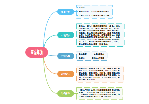
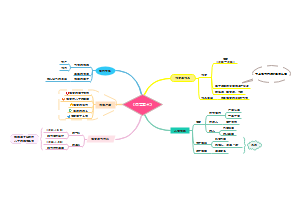
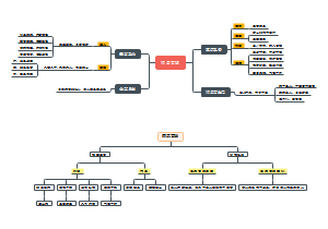
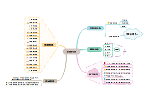
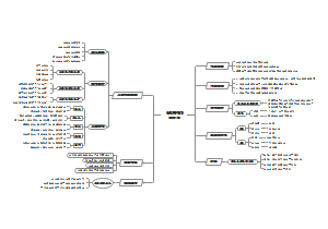
dentin repair
这是一篇关于dentin repair的思维导图,全篇用英语阐述,框架清晰,内容全面。适合英语写作以及练习英语课文阅读的同学。
编辑于2022-10-14 13:55:53 福建省- 英语作文
- dentin repair
- 相似推荐
- 大纲


dentin remineralization material
dentin hypersensitive
tooth paste
active tetracalcium phosphate/monetite mixtures
Method:Dentin de/remineralization cycling protocol consisted of demineralization in 1% citric acid at pH 4.6 with following remineralization with toothpastes and soaking in artificial saliva.
Results: The coatings on dentin surfaces was significantly thicker, had comparable remineralization potential as commercial calcium silicate/phosphate (SENSODYNE®) and magnesium aluminum silicate (Colgate®) toothpastes and significantly higher than control saliva (p < 0.02). Surface roughness was significantly lower after treatment with prepared and SENSODYNE® dentifirice (p < 0.05). had comparable properties with commercial fluoride calcium silicate/phosphate or magnesium aluminum silicate dentifrices.
Hydroxyapatite, Fluoroapatite, and Zn-Mg-hydroxyapatite Nanocrystals
All patients fulfilled the study requirements, and no adverse effects were registered. A reduction in DH was registered in 90%, 100%, and 50% of patients using nHAP, nZnMgHAP, and nFAP-containing toothpastes with effect sizes 2.52 (confidence interval [CI] 95%: 0.82, 4.14), 3.30 (CI 95%: 1.33, 5.20), and 1.44 (CI 95%: 0.09, 2.72), respectively. At 4 weeks, Schiff index scores decreased significantly in all groups compared to baseline.
fluoro calcium phosphosilicate, calcium sodium phosphosilicate, and strontium chloride hexahydrate containing dentifrice
Group 1––fluoro calcium phosphosilicate (BioMin™), Group 2––calcium sodium phosphosilicate (NovaMin®), and Group 3––strontium chloride hexahydrate. Clinical effectiveness (VAS), perceived sensation score (verbal rating scale [VRS]), participants’ subjective assessment (four-item questionnaire) and oral health-related quality of life (Oral Health Impact Profile-14 [OHIP-14]) questionnaire) were assessed.
At week 2, the percentage reductions in VAS (Group 1: 58.19%; Group 2: 49.18%; Group 3: 52.69%) and VRS (Group 1: 58.19%; Group 2: 47.16%; Group 3: 49.05%) scores were higher in Group 1 as compared with other groups. fluoro calcium phosphosilicate bioactive glass containing desensitizing dentifrice treatment may provide better treatment response for the treatment of DH because of its early onset of action in relieving hypersensitivity symptoms as compared with other dentifrices
four groups: toothpaste containing 10% nano-HAP (10%nano-HAP), 15% nano-HAP (15%nano-HAP), 10% nano-HAP supplemented with potassium nitrate (KNO3) (10%nano-HAPKN), or calcium sodium phosphosilicate (CSPS).
With either air or cold stimulus, VAS indicated a significant (P < 0.001) reduction from baseline DHS at each time point with all test toothpastes. Among the nano-HAP toothpastes, 15%nano-HAP and 10%nano-HAPKN were consistent in DHS reduction with both stimuli. With either stimuli, the CSPS did not significantly differ from 15%nano-HAP and 10%nano-HAPKN at any time point. Toothpaste containing nano-HAP (10 or 15%) alone or supplemented with KNO3 was as effective as CSPS for relief of DHS symptoms when used at least twice daily.
The desensitizing agent chosen in this study was calcium sodium phosphor silicate that is delivered in two vehicles – toothpaste and mouthwash.
Available in toothpaste and mouthwash delivery vehicle, this study was carried out to compare the effectiveness on dentinal hypersensitivity (using the Airblast test and Cold test) and on tooth remineralization (using DIAGNOdent pen) in a 4-week period.
Within-group comparison showed a significant reduction (P < 0.05) in the air blast score (toothpaste – 68.53% and mouth rinse – 48.52%), cold test score (toothpaste – 56.38% and mouth rinse – 38.87%), and DIAGNOdent score (toothpaste – 20.35% and mouth rinse – 9.49%). In-between group comparison showed no statistically significant difference (P > 0.05).Desensitizing mouthwash is as effective as toothpaste in reducing DH with a fair remineralization potential comparable with that of the toothpaste.
Nanohydroxyapatite and 8% Arginine Containing Toothpastes
The desensitizing paste containing arginine provided a statistically significant reduction in DH and so did the paste containing nHA. Mean increase in amperage value (reduction in DH) was higher for nHA based than the arginine containing dentifrice. This difference was not statistically significant showing that both toothpastes are equally effective. Conclusions: The findings of the present study encourage the use of Nano-hydroxyapatite and arginine containing dentifrice as an effective desensitizing agent providing relief from symptoms 5 minutes after application and after 1 and 4 weeks.
Group A toothpaste containing 8.0% arginine, calcium carbonate, and 1450 ppm fluoride as sodium monofluorophosphate; Group B toothpaste containing 8% strontium acetate and 1040 ppm fluoride as sodium fluoride; and Group C toothpaste containing 5% potassium nitrate and 917 ppm fluoride as sodium monofluorophosphate.
Group A resulted in more improvement in the reduction of sensitivity at 2, 4, and 8 weeks compared to the other groups. Group A toothpaste containing 8.0% arginine, calcium carbonate, and 1450 ppm fluoride as sodium monofluorophosphate significantly reduces dentin hypersensitivity and more effective than Group B and Group C toothpaste. Group A toothpaste is the latest new tool in the armament of the modern dentist.
Group 1 –5% NovaMin, Group 2 – Propolis, Group 3 – 5% potassium nitrate, and Group 4 – 8% arginine.
One hundred dentin slices were cut from the crown section of molars. Etching with 37% orthophosphoric acid was done to open the tubules. Scanning electron microscope (SEM) study was done to ensure that the tubules are opened.Samples were brushed for 2 min twice daily with a soft toothbrush for 15 days and were stored in distilled water. The samples were rinsed under running water to remove the toothpaste.
5% NovaMin group showed more completely occluded dentinal tubules when compared to other groups. The differences among all the groups were statistically significant (P ≤ 0.05). In the present study, all materials, NovaMin, Propolis, potassium nitrate, and arginine, were effective in occluding dentinal tubules but NovaMin appeared most proficient in occluding tubules, followed by arginine, potassium nitrate, and Propolis. Topical NovaMin is an upcoming agent demonstrating desensitization and remineralization properties.
eggshell-based toothpastes ith or without titanium dioxide nanoparticles (TNPs), coded “TNPs eggshell toothpaste [TNPsESTP]” or “eggshell toothpaste [ESTP],”
Mid-coronal dentin discs, from 28 human extracted molar teeth, etched with 37% phosphoric acid for 60 seconds to simulate the hypersensitive dentin, were randomly divided into four groups—G1: no treatment (negative control), G2: ESTP treated, G3: TNPsESTP treated, and G4: Biorepair treated (positive control). All treated discs were brushed for 2 weeks and 1 month using a toothbrush simulator at 40 mm/s. Dentinal tubules occlusion was studied using a cross-polarization optical coherence tomography (CP-OCT) and scanning electron microscopy (SEM).
From CP-OCT, a marked increase in surface reflectivity of dentin was observed after brushing with tested toothpastes. ESTP and NPsESTP showed higher or comparable grayscale values than Biorepair indicating increase in surface density of dentin. From SEM, at 2 weeks, ESTP showed comparable number of completely occluded dentinal tubules to Biorepair. TNPsESTP showed significantly lower numbers of CODT than Biorepair. At 1 month, the number of CODT was difficult to count for all treated groups. Both ESTP and TNPsESTP showed significantly higher numbers of partially occluded dentinal tubules than Biorepair.
a 0.454% stannous fluoride/5% sodium tripolyphosphate (STP) toothpaste
a SnF2/STP toothpaste was applied by brushing two selected sensitive teeth before 1 min whole-mouth brushing, compared to 1 min whole-mouth brushing only, with a negative control toothpaste. DH was assessed via evaporative (air) (Schiff scale) and tactile (Yeaple probe) stimuli after 7 and 14 d of twice-daily brushing.
In Study 1, the test treatment significantly reduced DH at 7/14 d versus baseline (7/14 d Schiff difference: −0.74 [−0.84,−0.65]/−1.39 [−1.54,−1.23]; tactile: 6.00 [4.88,7.13]/15.30 [13.34,17.26]) In Study 2, both treatments reduced DH compared to baseline by both measures, but there were no significant between-treatment differences. Toothpastes were generally well-tolerated. Previous studies and Study 1 support SnF2/5% STP toothpaste efficacy; Study 2 results may have been influenced by placebo/Hawthorne effects
a hydroxyapatite toothpaste containing a Polyol Germanium Complex with threonine
patients assigned to the GARDA SILK toothpaste; patients receiving the conventional fluoride toothpaste for comparison purposes, and patients asked to brush their teeth without toothpaste. The modified Quigley-Heine Plaque Index was assessed at the baseline and after treatment. Sensitivity was assessed at baseline, 3, 7 and 14 days using the air stimulus (Schiff Index). The impact of dentine hypersensitivity on the quality of life was assessed before and after the treatment with the Russian version of the Dentine Hypersensitivity Experience Questionnaire (DHEQ).
After 14 days of treatment, the mean Plaque Index scores in Groups 1 and 2 did not exceed 1 and averaged 0.72 ± 0.249 and 0.86 ± 0.213, respectively. In Group 3, the mean Plaque Index was significantly higher (1.04 ± 0.070, p < 0.05). The mean Schiff sensitivity scores in Group 1 decreased to 2.45 ± 0.42 at 3 days, 1.84 ± 0.26 at 7 days, and 1.02 ± 0.34 at 14 days. Group 2 exhibited smaller reductions in the Schiff index at all time points. In this group, Schiff scores dropped to 2.67 ± 0.28 after 3 days, 2.34 ± 0.44 after 7 days, and 1.93 ± 0.36 after 14 days. The GARDA SILK hydroxyapatite toothpaste with a threonine-containing Polyol Germanium Complex (PGC) is effective in maintaining good oral hygiene, reducing dentine hypersensitivity, a
paste and gel
plain nano-hydroxyapatite paste
Group 1: NanoXIM® n-HA paste (15% plain n-HA, FLUIDINOVA, S. A., Moreira da Maia, Portugal) Group 2: Fluoraphat Pro® paste (Sodium fluoride (NaF) 5%, Promedica Dental Material GmbH, Neumünster, Germany) Group 3: : Placebo (Glycerin, water, Ivory Med Company, Riyadh, Kingdom of Saudi Arabia) The patient's response to dentin hypersensitivity (DH) was evaluated at baseline (T0), immediately after application (T1), and after 1 week (T2). Tactile and cold air sensitivity (CAS) stimulus tests and visual analog scale subjective tests were used to measure the patient's response to DH.
All three groups showed a statistically significant reduction in tactile sensitivity (TS), cold air sensitivity, and visual analog scale scores from T0–T1 and T0–T2 (P < 0.005). However, from T1–T2, there was no significant reduction in the TS, cold air sensitivity, and visual analog scale scores of the groups (P > 0.005) except for Group 1, which showed a significant reduction (P = 0.033) in visual analog scale scores from T1–T2. However, the intergroup comparison at T2 demonstrated a significant difference in tactile scores (P = 0.005), CAS (P < 0.001), and visual analog scale scores (P < 0.001). There was no incidence of treatment-related adverse events with the study products. n-HA paste was the most effective desensitizing paste compared to fluoride and placebo pastes. The single application of the n-HA paste demonstrated a significant reduction in visual analog scale scores after 1-week.
Group 1—Teethmate Desensitizer (TD) (Noritake Dental Inc., Tokyo, Japan), a a pure tetracalcium phosphate (TTCP) and dicalcium phosphate dihydrate (DCPD) calcium phosphate powder and water liquid; Group 2—Dentin Desensitizer (DD) (Ghimas, Casalecchio di Reno, Bologna, Italy), a premixed n-HAP alcohol-based gel, also containing 30% potassium oxalate; Group 3—Bite&White ExSense (BWE) (Cavex Holland, Haarlem, Netherlands), a premixed n-HAP water-based gel, also containing potassium nitrate
All the materials decreased DH after 24 weeks in comparison to Pre-1. However, the TTCP/DCPD cement showed the greatest statistical efficiency.
Polymethylmetacrylate-based nanoparticles (NPs) were doxycycline (D), calcium, or zinc loaded.
Dentin treated with Zn-NPs attained the highest nanomechanical properties, mineralization, and crystallinity among groups. Nanoroughness was lower in Zn-treated surfaces in comparison to dentin treated with undoped gels. Dentin treated with Ca-NPs created the minimal calcification at the surface and showed the lowest Young’s modulus at peritubular dentin. Intertubular dentin appeared remineralized. Dentinal tubules were empty in samples treated with D-NPs, partially occluded in cervical dentin treated with undoped NPs and Ca-NPs, and mineral covered when specimens were treated with Zn-NPs. Zn-loaded NPs permit functional remineralization of eroded cervical dentin. Based on the tested nanomechanical and chemical properties, Zn-based nanogels are suitable for dentin remineralization.
amorphous calcium magnesium phosphate (ACMP) microspheres
Transmission electron microscopy (TEM) and scanning electron microscopy (SEM) showed that crystallization was initiated on the peritubular dentin (PTD) with undirected crystal growth leading to the formation of a porous material. We additionally investigated the effects from using a fluoride toothpaste to potentially improve the remineralization and anti-cariogenic properties of the ACMP microspheres. Energy dispersive x-ray spectroscopy (EDX) using TEM in scanning mode (STEM) showed that fluoride incorporation resulted in an increase in aspect ratio of the crystals, crystal growth directed towards the center of the tubule lumen and densification of the mineralized material.
Varnish
group 1: Sodium fluoride 5% varnish was applied (positive control), Group 2: No treatment (negative control), Group 3: Treated with Remin Pro (contains hydroxyapatite and fluoride), Group 4: Treated with MI paste (contains casein phosphopeptide-amorphous calcium phosphate [CPP-ACP]) and Group 5: Treated with GC tooth mousse (contains CPP-ACP). Group 1 (positive control): Sodium fluoride (NaF) 5% varnish was applied Group 2 (negative control): No treatment material was applied Group 3: Specimens were treated with Remin Pro (VOCO, Cuxhaven, Germany) Group 4: Specimens were treated with MI paste (GC, Melbourne, Australia) Group 5: Specimens were treated with GC tooth mousse. (GC, Melbourne, Australia) SEM images were obtained and mean tubular diameter was measured in each group.
Statistically significant difference was observed between Group 2 (negative control) and other four groups (P < 0.05). There was no significant difference between Groups 1, 3, 4, and 5 (P > 0.05). Under the limitations of the present in vitro study, it can be concluded that the application of a CPP-ACP paste as well as a paste which contains fluoride is effective on reduction of dentin permeability.
a calcium-fluoride-forming agent (Tiefenfluorid®, Humanchemie GmbH, Alfeld, Germany) with that of a fluoride varnish (EnamelastTM, Ultradent Inc., Cologne, Germany)
Within the limits of the present study, Tiefenfluorid® was more effective than EnamelastTM against DH in that it provided long-lasting results, with a significant improvement still detected at the latest 6-month follow-up
a novel zinc-containing desensitizer CAREDYNE Shield
the CAREDYNE Shield group (intervention group) and the Nanoseal group (control group). The pain intensity in response to air stimuli, gingival condition, and oral hygiene status of CDH teeth were assessed before and at 4 weeks after treatment.
A significant reduction of pain in response to air stimuli was observed in both groups; however, no significant difference was observed between the groups. This study showed that CAREDYNE Shield is effective for CDH and its effectiveness is similar to Nanoseal.
Ca/PO4−/F− varnish (Clinpro White Varnish) and ionomeric sealant (IS) (Clinpro XT Varnish) with a placebo.
IS showed the highest VAS value difference between baseline and 4 weeks (mean ± SD: 3.7 ± 2.2), differing significantly from placebo (2.3 ± 1.7) and Ca/PO4/F− varnish (2.6 ± 2.0). Ca/PO4−/F− varnish did not differ from placebo. In all time intervals, VAS values of all groups were significantly lower than the baseline value, without differences among groups. For the long-term differences in VAS values (3 and 6 months), the IS exhibited significantly higher values than Ca/PO4−/F− varnish. The IS was the most efficacious product for reducing DH, whereas Ca/PO4−/F− varnish did not differ from placebo.
experimental materials
pure nano-hydroxyapatite (nHAP) and 1%, 2%, and 3% F¯ doped nano-HAp
All of the nano-HAp materials used in this study built up an effective covering layer on the dentin surfaces even with plugs in tubules. It was found that this layer had also a resistance to degradation. None of the evaluated nano-HAp types were have toxicity. Fluoride doping showed a positive effect on physical and chemical stability until a critical value of 1% F−.
spherical particles of amorphous calcium magnesium phosphate (180–440 nm in diameter)
A degradation study of the particles in Tris-HCl buffer showed that the particles continuously released Ca2+, Mg2+, and phosphate, and XRD analysis revealed the formation of hydroxyapatite (HA) after 1 week. Application of the particles followed by incubation in artificial saliva resulted in occlusion of exposed tubules, and examination with SEM showed that the particles could penetrate the tubules down to 100 μm from the dentin surface. Transformation of the particles into nanocrystalline HA-structures (nanoHA) was initiated at the dentin surface within 12 h of application, and tubule penetration of the particles, accompanied by further ion release and diffusion of ions, resulted in deep intratubular occlusion in the majority of the tubules within 3 days from application. NanoHA was tightly adhered to the tubule walls, filling the entire tubule volume after 7 days. The results of this study demonstrate the mode of action of the amorphous calcium magnesium phosphate particles in occluding exposed dentin tubules.
pharmaceutical composition developed by the authors based on hydroxyapatite (HAp).
The chemical composition of the dentin surfaces was analyzed using scanning electron microscopy (SEM) equipped with an energy-dispersive X-ray spectroscope (EDS), Fourier-transform infrared (FTIR) and Raman spectra techniques. The specimens were immersed in an artificial saliva solution for 24 h, 48 h and 7 days to assess the durability of the layers and the tubule-obliteration effectiveness.
All the test groups showed some degree of dentinal tubule occlusion or a covering layer, but the HAp-based composition proved to be the longest-lasting. It was concluded that the developed pharmaceutical composition creates a coating on the dentin surface built of hydroxyapatite crystals sized 10-20 μm, which are likely to constitute a reservoir of calcium and phosphate ions, as well as smaller crystals (0.2-0.3 μm) that occlude dentinal tubules.
spherical mesoporous bioactive glass nanoparticle (MBGN) and non-porous bioactive glass nanoparticle (BGN)
Group 1, no treatment; Group 2, Dense BGN; Group 3, MBGN. Then, four discs per group were treated with 6wt.% citric acid challenge to determine the acidic resistance. The effects on dentinal tubule occlusion were observed by FESEM. The microtensile bond strength (MTBS) was also measured.
Dense BGN and MBGN occluded the dentinal tubule before and after acid challenge. However, only MBGN formed a membrane-like layer and showed hydroxyapatite formation after soaking SBF solution. There were no significant differences in MTBS among dense BGN, MBGN (P>0.05).
The porous bio-calcium carbonate-silica (BCCS) (1) BCCS mixed with H3PO4; (2) BCCS mixed with KH2PO4; (3) Seal & Protect® was used as a comparison group
Sealing efficacy was evaluated by measuring the depths and percentages of precipitate occlusion in dentinal tubules with SEM.
The N2 adsorption–desorption isotherm of the BCCS demonstrated a pore size of around 15.0 nm and a surface area of 61 m2g-1. From the results of occlusion percentage and depth, the BCCS treated with H3PO4 or KH2PO4 demonstrated promising sealing efficacy than the commercial product.
fluoride varnish and self-etching adhesive, and the presence of nanofibers: C (self-etching adhesive Clearfil SE Bond), CN (Clearfil SE Bond with 1% nanofiber), D (Duraphat varnish), and DN (Duraphat varnish with 1% nanofiber).
The C group showed the lowest hydraulic conductance (Lp%) (89.33), while the DN group showed the highest Lp% (116.06). No statistical significance was observed in the Lp% values in all groups after the treatment and 6% citric acid challenge (p > 0.239). In the images, the CN group presented a higher superficial and intratubular deposition. In addition, this group presented a more homogeneous dentin surface and wide occlusion of dentinal tubules than the other treatments. Despite there being no statistical differences among the treatments employed, the images showed that the CN group presented a higher surface and intratubular deposition compared to the other treatments, even after the acid challenge.
hydroxyl apatite functionalized calcium carbonate (FCC) particles
Dentine specimens a) Cut surface with smear layer; b) EDTA (smear layer removed with 17% EDTA for 1 min); and c) Grit blasted functionalized calcium carbonate (FCC) with and air pressure of 280 kPa. Biomineralization of specimens was carried out in a simulated body fluid (SBF).
FCC particles showed penetration into the dentinal tubules by breakage of their original particle shape and size. EDTA treated surface had higher number and larger size tubules than those with smear layer or grit blasted (p < 0.005). SEM-EDX analysis revealed mineral precipitation of calcium phosphate on the SBF immersed dentin specimens. XRD analysis showed typical crystal structure of hydroxyl apatite for the biomineralized surface layer on dentine. Grit blasted FCC particles initially occluded effectively the opened dentinal tubules and biomineralization occurred in tubules primarily occluded by the FCC particles. However, in the optimal in vitro conditions in SBF, no difference between biomineralization was found between the grit blasted surface and the control surface.
Laser related
a bioactive glass (BG) paste (BG/Ac) irradiated or not with high-power lasers.
application of BG/Ac by itself caused some obstructions of dentinal tubules. when BG/Ac paste was irradiated with lasers, a sequence of surface reactions between glass and dentin interface led to the formation of an amorphous hydroxyapatite layer, similar to that of an inorganic component of the normal dentin. Moreover, BG/Ac was able to prevent the formation of cracks and degradation of collagen fibers caused by CO2 irradiation.
combined diode laser and GLUMA bonding therapy with combined diode laser and 5% sodium fluoride varnish(clinical experiment)
30th day after the intervention,The combined treatment with GLUMA bonding and the 660 nm diode laser is effective in reducing DH and this is more effective than GLUMA bonding alone in the long term. However, it does not have a significant advantage over the combined varnish-laser method, but it seems that due to its ease of use, it can be a suitable alternative to the varnish-laser method.
Ca2+/PO43−@mesoporous silica nanoparticles (MSNs)
The shape and structure of the MSNs were examined using transmission electron microscopy (TEM) and scanning electron microscopy (SEM). The surface morphology and chemical compositions of Ca2+@MSNs/PO43−@MSNs and Ca2+/PO43−@MSNs were examined using SEM and X-ray fluorescence (XRF). The element distribution of Ca2+/PO43−@MSNs was detected using energy dispersive spectrometer (EDS). The sustained release ability of Ca2+@MSNs/PO43−@MSNs was detected using inductively coupled plasma atomic emission spectrometry (ICP-AES). The efficacy of Ca2+/PO43−@MSNs on dentinal tubule sealing was evaluated using SEM, and the results were analyzed by Image-Pro software to determine the best water-powder ratio. We also compared the sealing efficacy between Ca2+/PO43−@MSNs and NovaMin, which is currently used in clinics, under the simulated conditions of oral acidic corrosion and mechanical friction.
Ca2+/PO43−@MSNs are a new type of tubule-occluding material with sustained release properties. The ratio of Ca2+@MSNs: PO43−@MSNs: H2O =0.015 g: 0.015 g: 150 µL exhibited an excellent sealing effect on dentinal tubules as well as resistance to oral acid corrosion and daily oral friction. The novel dental material Ca2+/PO43−@MSNs demonstrates potential long-term effectiveness in sealing dentinal tubules and reducing dentin sensitivity, which is one of the most important problems in dental clinics.
dentin filling materials Caries
top-down remineralization
An experimental adhesive resin was formulated with BisGMA, HEMA (66.66:33.33 wt%), and a photoinitiator system. α-TCP was added at 2 wt% into this base resin
The experimental adhesive resin with α-TCP showed higher initial Knoop hardness than the control group (p < 0.05). There was no difference regarding the softening in solvent (p > 0.05) and the DC (p > 0.05). The polymerization rate of α-TCP group was higher than the control. There was no difference for μ-TBS between groups (p > 0.05). The hardness of CAD increased in 100 and 200 μm from the adhesive interface after three months (p < 0.05), and both groups indicated higher mineral content on CAD by micro-Raman spectroscopy. The α-TCP addition did not affect the dental adhesives’ properties and did not change the mineral deposition and μ-TBS.
nanoparticles of amorphous calcium phosphate (NACP) and dimethylaminohexadecyl methacrylate (DMAHDM)
The in vitro experiment results showed that the NACP + DMAHDM adhesive effectively achieved acid neutralization, decreased biofilm colony-forming unit (CFU) count, decreased biofilm lactic acid production, and increased biofilm calcium and phosphate content. The NACP + DMAHDM adhesive group had higher remineralization value than the NACP or DMAHDM alone adhesive group. The NACP + DMAHDM adhesive was effective in remineralizing dentin lesion in a biofilm model. It is promising to use NACP + DMAHDM adhesive to protect bonded interface, inhibit secondary caries, and prolong the longevity of restoration.
an adhesive containing nanoparticles of amorphous calcium phosphate (NACP)
The NACP adhesive achieved acid neutralization, decreased biofilm CFU count, decreased biofilm lactic acid production, and increased biofilm calcium and phosphate content (P < 0.05). The NACP adhesive group had higher remineralization value than the commercial fluoride-releasing adhesive group (P < 0.05).
a dual-functional nanocomposite with antibiofilm and remineralization properties was designed by combining zwitterionic poly(carboxybetaine acrylamide) (PCBAA) and ACP.
The resulting nanocomposite was stable in solution for at least 3 days without any aggregation. The PCBAA/ACP nanocomposite exerted a significant inhibitory effect on the adhesion and biofilm formation of Streptococcus mutans and exhibited bactericidal activities under acidic conditions resulting from bacteria. Moreover, compared with fluoride, this nanocomposite demonstrated superior effects in promoting the remineralization of demineralized enamel and the occlusion of exposed dentinal tubules in vivo and in vitro. The present work provides a theoretical and experimental basis for the use of the PCBAA/ACP nanocomposite as a potential dual-functional agent for arresting and preventing caries.
NACP was mixed into Scotchbond Multi-Purpose (SBMP, 3M, St. Paul, MN, USA) adhesive at mass fraction of 40%, and DMAHDM was mixed into the adhesive at mass fraction of 5%
the NACP + DMAHDM adhesive effectively achieved acid neutralization, decreased biofilm colony-forming unit (CFU) count, decreased biofilm lactic acid production, and increased biofilm calcium and phosphate content. The NACP + DMAHDM adhesive group had higher remineralization value than the NACP or DMAHDM alone adhesive group.
3% nanoparticles of amorphous calcium phosphate (NACP) and 20% dimethylaminohexadecyl methacrylate (DMAHDM) 55.8 wt % urethane dimethacrylate (UDMA) and 44.2 wt % triethylene glycol divinylbenzyl ether (TEG-DVBE)
Adding DMAHDM and NACP into low shrinkage-stress composite did not compromise the flexural strength. The low-shrinkage-stress composite with DMAHDM achieved substantial reductions in biofilm colony-forming units (CFU), lactic acid production, and biofilm biomass (p < 0.05). The low-shrinkage-stress DMAHDM+NACP composite exhibited no significant difference in antibacterial performance before and after 3 months of aging, demonstrating long-term antibacterial activity. Under S. mutans biofilm acidic attack, dentin hardness (GPa) was 0.24 ± 0.04 for commercial control, and 0.23 ± 0.03 for experimental control, but significantly higher at 0.34 ± 0.03 for DMAHDM+NACP group (p < 0.05). At an instrumental compliance of 0.33 μm/N, the polymerization shrinkage stress of the new composite was 36% lower than that of a traditional composite (p < 0.05). The triple strategy of antibacterial, remineralization and lower shrinkage-stress has great potential to inhibit recurrent caries and increase restoration longevity. Polymerization shrinkage stress, masticatory load over time as well as biochemical degradation can lead to marginal failure and secondary caries. The present study developed a new low-shrinkage-stress, antibacterial and remineralizing dental nanocomposite. Polymerization shrinkage stress was greatly reduced, biofilm acid production was inhibited, and tooth dentin mineral and hardness were preserved.
The UV-experimental composite had a whitish light color, and the addition of NACP resulted in a slight grayish shade. Further study is needed to investigate the effect of the addition of colorants on the shades on the new composite. Future studies should investigate the depth of cure, viscosity, and flow properties of this new material.
EXP, an experimental resin-based bioactive material consisting of a self-etch primer and an adhesive containing a fluoride-doped bioglass; GIC, a glass ionomer cement (Riva LC); MTA, Mineral Trioxide Aggregate (ProRoot MTA); BIO, a calcium silicate cement (Biodentine). simulated caries lesion: (D1) Shallow chemically-induced caries, (D2) deep chemically-induced caries, (D3) deep bacterially-induced caries.
All four restorative materials induced mineral gains regardless of the protocol for caries lesion, without significant differences between materials. Microhardness significantly increased in the groups BIO and MTA, but not GIC; EXP only provided hardness gains in D3-lesions. Fluorescence and confocal microscopy confirmed these results. There was a clear “top-down” remineralization in the groups BIO and MTA, and “bottom-up” intrafibrillar collagen remineralization in EXP.
dentin bonding agents containing different ratios of nano-sized hydroxyapatite fillers (HA)
The maximum mean value of micro-tensile bond strength (μTBS) was in the 7% HA group, while the minimum mean value of μTBS was observed in the control group. 7% HA group was statistically significant and higher than other groups while there were no significant differences between the control, 2% HA, and 5% HA groups. According to SEM analysis, fracture analysis revealed that the mixed fracture type was seen more often than the other fracture types. The particle size and amount of HA fillers added to the adhesive resin seem to affect the success of the bond strength to the dentin. Adding different ratio nano-sized HA fillers to the adhesive resin contributed positively to the immediate μTBS values in the dentin.
ZrO2 particles Experimental adhesive resins
The addition of ZrO2 did not influence the radiopacity. The addition of 4.8% and 9.1 wt.% ZrO2 showed higher initial hardness with increased softening in solvent (P < 0.05) and promoted mineral deposition at the dentin interface. degree of conversion (DC) was significantly increased in the group with 1% ZrO2 (P < 0.05). The μTBS test showed difference on the group with 9.1 wt.% of ZrO2, with a significant reduction after aging.
buttom-up remineralization
calcium silicate based and the second is calcium hydroxide based materials. biomimetic analogs (poly-acrylic acid and sodium tri-meta-phosphate)
Calcium silicate-based cement group with biomimetic analogs showed the highest statistically significant calcium and phosphorous wt% in addition to highest surface hardness values after 12 weeks of storage. Demineralized dentin ground sections showed increase in light zones after total period of storage. Calcium silicate-based cement showed the best ability to enrich the artificial carious dentin with ions for remineralization. Using biomimetic analogs had a significant impact on demineralized dentin surface hardness improvement.
Novel ionomeric cement compositions based on bioglass 45S5 and pAsp mixtures, as well as conditioning solutions (conditioner) containing 5 mg/mL pAsp,
This study showed that functional remineralization of artificial lesions using PILP-releasing restoratives occurred, indicated by an increase of the elastic modulus in shallow lesions and in the middle zone of deep artificial lesions. The mechanical improvement was significant when compared to glass ionomer cement (RMGIC) restoration without pAsp (P < 0.05). Nonetheless, recovery across artificial lesions was most significant when specimens were immersed into PILP-solution with restorative (P < 0.01). Furthermore, natural lesions increased in mineral volume content to a higher degree when the restorative treatment included the PILP-method (P < 0.05). However, none of the natural lesions recovered to full mineral degree regardless of the treatments.
a tripolyphosphate (TPP) “templating analog” and a poly(acrylic acid) (PAA) or poly(aspartic acid) (pAsp) “sequestration analog,” the latter of which generates the polymer-induced liquid-precursor (PILP) mineralization process
No mineralization was observed in any of the PAA groups. In both the pAsp and no polymer groups, TPP inhibited mineralization on the surfaces of the specimens but promoted mineralization within the interiors. Pre-treatment with TPP enhanced overall mineralization of the pAsp group. However, when analysed via TEM, regions with little mineral were still present.
casein phosphopeptide–amorphous calcium phosphate combined with sodium tripolyphosphate
A (the CPP–ACP group), B (the CPP–ACP + TPP combination group), C (the artificial saliva group), D (the negative control group), and E (the positive control group). Dentin slice samples from groups A, B and C were remineralized and the remineralization effect was evaluated using scanning electron microscopy (SEM), transmission electron microscopy (TEM), energy-dispersive X-ray spectroscopy (EDX), attenuated total reflection–Fourier transform infrared spectroscopy (ATR–FTIR) and X-ray diffraction (XRD).
Treatment with CPP–ACP combined with TPP occluded the dentinal tubules and resulted in remineralization of collagen fibrils. The hydroxyapatite crystals formed via remineralization were found to closely resemble the natural dentin components.
carboxylated polyamidoamine dendrimer/amorphous calcium phosphate nanocomposite (PAMAM–COOH/ACP)
PAMAM–COOH/ACP showed superior remineralization ability of human dentin type I collagen fibrils, especially the intrafibrillar remineralization.
calcium phosphate polymer-induced liquid precursor (Ca/P-PILP): a double NCP analogues process-directing agent of PAA-PASP
Solely PAA-PASP solution and solely saturated Ca/P solution can’t achieve dentin collagen remineralization. Increased concentration of Ca/P-PILP and prolonged remineralization time can enhance the biomimetic remineralization intensity of demineralized dentin collagen. After treating with high concentration Ca/P-PILP, a 150 ± 50 μm thick layer of demineralized artificial caries dentin lesion was not fully remineralized, and the biomimetic remineralization intensity reached up to 88.0%. Furthermore, a better bonding interfacial integrity with less microgap and increased bond strength at both baseline level and aging level were observed when artificial caries dentin lesion was biomimetically remineralized with high concentration Ca/P-PILP. Biomimetic remineralization of demineralized caries dentin lesion promotes its clinical properties for resin composited adhesive restoration.
the effect of conditioning solutions containing DL-aspartic amino (Asp) on dentine remineralization induced by bioactive glass 45S5 (BAG)
Polyacrylic acid (5%) and sodium trimetaphosphate (5%) were added to the primer and monocalcium phosphate monohydrate (9%), beta-tricalcium phosphate (10.5%), and calcium hydroxide (0.5%) were added to the adhesive
EDS identified the presence of isolated calcium phosphate nanoparticles in the demineralized region; however, the SAED analysis did not show any evidences of hydroxyapatite (HA) neoformation in SD and CAD. The biomimetic analog-based adhesive system inhibited the activities of dentin proteases immediately after treatment. Additionally, the proteolytic activity on the affected dentin resembled that of the SD. In conclusion, no HA formed in the demineralized SD and CAD although there were calcium and phosphate deposits. The experimental adhesive system inhibited dentin proteases.
Both BAG and Asp-BAG groups significantly reduced dentine permeability and formed enamel-like apatite layers on dentine surface. For the mineralization of BAG, Asp showed inhibition effect. The 7-day mineral matrix area ratio in BAG group (12.54 ± 2.29) was lower than the value in the Asp-BAG group (17.77 ± 2.27) (p < 0.05) and the Raman intensity (RI%) in Asp-BAG Group (1.49 ± 0.26) was also significantly higher than that of BAG group (1.34 ± 0.14) (p < 0.05). According to permeability test, the apatite layer in BAG group and Asp-BAG group effectively occluded the dentinal tubules (p < 0.05) and had certain acidic resistance (p > 0.05). Furthermore, adsorbed acidic amino acid on hydroxyapatite (HAP) altered the crystal to increase into a larger size in diameter during crystal growth. The study demonstrated that a superior remineralization efficacy of BAG with Asp pretreatment on dentine.
the phosphate-terminated polyamidoamine dendrimer (PAMAM-PO3H2) carboxylterminated dendrimers (PAMAM-COOH)
the surfaces of human dentin were covered with regenerated crystals and the dentinal tubules were occluded by PAMAM-PO3H2 and PAMAM-COOH. In summary, the combination of PAMAM-PO3H2 and PAMAM-COOH may be another feasible therapeutic method for the treatment of dentin caries and dentin hypersensitivity.
carboxymethyl chitosan (CMC)
TEM, SEM, and SIM images showed that CMC had a positive effect on stabilizing amorphous calcium phosphate (ACP) and promoting intrafibrillar mineralization, while extrafibrillar mineralization was formed without CMC. Furthermore, hardness evaluation and µTBS proved that CMC significantly increased dentin hardness and bond strength. CLSM indicated that CMC could create a significantly better bond interfacial integrity with less of a micro-gap in ACAD. CMC possessed the ability to promote intrafibrillar mineralization and remineralization in demineralized caries dentin lesions, as well as improve bond performance, which implied its potential in carious dentin demineralization or dentin hypersensitivity and possibly even as a possible material for indirect pulp-capping, to deal with deep caries.
carboxymethyl chitosan (CMC)
Transmission electron microscopy and selected area electron diffraction conducted on reconstituted two-dimensional collagen showed typical deposition of needle-like hydroxyapatite crystals within collagen fibrils through CMC-induced biomimetic mineralization. The Vickers hardness test revealed significant improvement (P < 0.001) of the hardness of ACAD treated with CMC-containing experimental resins. Confocal laser scanning microscopy showed reduced dentin permeability and defect sites after biomimetic mineralization. On microtensile bond strength testing, the CMC-remineralized ACAD had better bonding with resin than ACAD and traditionally remineralized ACAD in both self-etch and etch-and-rinse bonding modes (P < 0.001). In conclusion, CMC is efficient in directing the biomimetic mineralization of collagen fibrils. The experimental resins containing CMC can induce dentin biomimetic remineralization and improve the bonding performance of ACAD.
collagen/hydroxyapatite (Col/Hap) nanocomposite together with grape seed extract (GSE; 6.5%)
Applied Col/Hap (30/70%) together with GSE (6.5%) gave the significantly highest μTBS (25.04 ± 5.47 and 25.53 ± 7.64 MPa, for 10min and 1 h application times, respectively). After thermocycling for 10,000 cycles at 5 and 55 °C, μTBS for all protocols and both application times substantially decreased especially for the two control groups. Using the suggested dentin pre-treatment protocols, in chair-side, may possibly enhance the bond strength to the deeper and remineralizable carious zone (DRCZ) in dentin and its durability.
new types of calcium phosphate ion clusters (CPICs) CPICs were synthesized as specified by Shao C. et al. [6]. Two solutions were prepared for the synthesis of CPICs. Solution A, 0.20 g of CaCl2·2H2O and 3.8 mL of triethylamine(TEA) were added into 80 mL of ethanol and then ultrasonicated (BRANSON, Danbury, CT, USA) for five min. Solution B, 70 µL of H3PO4 was added into 20 mL of ethanol and stirred thoroughly. Solution B was dropped into solution A, with slight agitation, and then CPICs (2 mg/mL) formed in the solution.
In the SEM imaging, with a rising concentration of CPICs, the degree of remineralization of dentin increased significantly. The metastable Ca-P treated specimens showed a similar level of remineralization as the 1 mg/mL CPICs treated specimens. The TEM imaging also revealed that dentin remineralization occurs in a CPICs concentration-dependent manner between the demineralized dentin and the resin layer. Furthermore, the results of micro-tensile bond strength showed the same trend as the results confirmed by SEM and TEM. We demonstrated that a 1 min pretreatment of CPICs or metastable Ca-P in etched dentin collagen fibril can achieve biomimetic remineralization and increase micro-tensile bond strength.
Nacre
The results demonstrated that the application of “seawater-like” mineralizing precursor medium effectively occluded dentinal tubules, reduced dentine permeability, increased surface microhardness, provided certain acid-resistant stability and possessed favorable in-vitro biocompatibility. In addition, the dynamic procedure of hierarchical intrafibrillar nanocrystalline assembly was observed, which offered a clue to uncover its remineralizing mechanism. It is attainable to realize the biomimetic remineralization of human dentine via the “bottom-up” concept inspired by nacre structure duplication, suggesting great potential for providing dentists a therapeutic strategy to counter dentine hypersensitivity in the future.
polydopamine (PDA) polyacrylic acid
dentine was successfully repaired using the biomolecule polydopamine (PDA), and the remineralized dentine exhibited mechanical properties comparable to those of natural dentine. Detailed analyses of the collagen mineralization process facilitated by PDA showed that PDA can promote intrafibrillar mineralization with a decreased heterogeneous nucleation barrier for hydroxyapatite (HAP) by reducing the interfacial energy between collagen fibrils and amorphous calcium phosphate (ACP), resulting in the conversion of an increasing amount of nanoprecursors into collagen fibrils. The present work highlights the importance of interfacial control in dentine remineralization and provides profound insight into the regulatory effect of biomolecules in collagen mineralization as well as the clinical application of dentine restoration.
small leucine-rich proteoglycan (SLRP) proteins DCN core, DCN + GAGs, BGN core, BGN + GAGs.
MTBS test presented a mean of 51.4 ± 9.1 MPa in control with no statistically significant difference to DCN core (47.6 ± 8.3) and BGN core (48.3 ± 6.5). The full proteoglycan groups DCN + GAGs (27.4 ± 4.5) and BGN + GAGs (36.4 ± 13.6) showed decreased MTBS compared to control (p < 0.001). At 6 months, control or core-treated samples did not have a statistically significant difference in MTBS. However, SLRPs with GAGs showed statistically significant improvement of bonding (62.5 ± 6.0 for DCN and 52.8 ± 8.1 for BGN, p < 0.001) compared to their baseline values. SEM showed that GAGs seem to favor water retention but overtime help remineralization. TEM of demineralized dentin indicated a larger collagen fibril diameter pattern of samples treated with core proteins compared to control and a smaller diameter with DCN + GAGs in water with evidence of mineralization with DCN + GAGS, BGN core and BGN + GAGs. In conclusion, core proteins seem not to affect dentin adhesion significantly but the presence of GAGs can be detrimental to immediate bonding. However, after ageing of samples, full proteoglycans, particularly DCN, can significantly improve bonding overtime while promoting remineralization which can prove to be clinically beneficial.
10 & 20% Strontium doped nano-hydroxyapatite (nHAp) with or without NCP analogue-chitosan.
It was observed that 20% Sr-nHAp paste and the experimental formulations with Sr-nHAp and NCP analogue chitosan had the potential to remineralise the partially demineralised dentin specimens as observed by both increase in the mineral content noted in the SEM-EDS analysis and increased mechanical properties analysed by nanoindentation. Within the limitations of the study it was observed that Chitosan enhanced remineralisation potential of 10 & 20% Sr-nHAP and improved mechanical properties of partially demineralised dentin and hence has potential application as a remineralising agent and in vital pulp therapy procedures.
Comparation above
EXP, an experimental resin-based bioactive material consisting of a self-etch primer and an adhesive containing a fluoride-doped bioglass; GIC, a glass ionomer cement (Riva LC); MTA, Mineral Trioxide Aggregate (ProRoot MTA); BIO, a calcium silicate cement (Biodentine). Specimens were mounted in a dual-chamber device to simulate the exposure to pulpal pressure and oral fluids. After 3 months, mineral and mechanical gains were assessed using transverse microradiography (vol% × μm) and microhardness measurements (VHN). Characterization using confocal microscopy and transmission electron microscopy (TEM) was also performed.
All four restorative materials induced mineral gains regardless of the protocol for caries lesion, without significant differences between materials. Microhardness significantly increased in the groups BIO and MTA, but not GIC; EXP only provided hardness gains in D3-lesions. Fluorescence and confocal microscopy confirmed these results. There was a clear “top-down” remineralization in the groups BIO and MTA, and “bottom-up” intrafibrillar collagen remineralization in EXP. Mineral gains did not always translate into hardness gains. Biodentine and MTA induced evident mineral precipitation, but intra/inter-fibrillar collagen mineral infiltration was only provided by biomimetic remineralisation via the use of the experimental adhesive.
indirect pulp capping materials
two hydraulic calcium-silicate cements (Biodentine and TheraCal LC)
Demineralized dentin, next to Biodentine, showed statistically higher intensities of Ca and P wt% after the three periods of incubation (p< 0.05). Surface mapping of both tested cements and their adjacent demineralized dentin showed increase in overall distribution of previous ions. SEM of subsurface layer under both materials showed filling of most intra-tubular areas with rod-like mineralized structure without significant difference. Biodentine has a higher ability to enrich the artificial carious dentin with significantly higher mineral contents available for remineralization. Both pulp-capping materials have significantly induced remineralization of demineralized dentin beneath them after total period of incubation.
Surface pre-reacted glass (S-PRG) fillers incorporating LiCl
We show that treatment of human dental pulp stem cells with eluates from S-PRG/LiCl combination cements leads to an upregulation in cell migration, differentiation, and mineralization in vitro. In pulp-capping animal trials, we found that S-PRG/LiCl cements could induce tertiary dentin formation 28-days post-capping. At 7 days post-capping, we identified both β-catenin and Axin2 expression using immunofluorescence, indicative of Wnt/β-catenin signaling activity. In conclusion, S-PRG/LiCl cement is highly effective in promoting human dental pulp stem cells profiles and in enhancing reparative dentin formation in rat teeth through activation of the Wnt/β-catenin canonical signaling pathway.
pulp-capping agents: (I) hard-setting calcium hydroxide (Dycal) (control group) (n = 36), (II) bioactive tricalcium silicate (Biodentine) (n = 37), and (III) resin-based tricalcium silicate (TheraCal LC) (n = 36)
At the end of the 24-month follow-up period, the clinical and radiographic success rates for Dycal, Biodentine, and TheraCal LC were 100%, 100%, and 93.3%, respectively, and there was no significant difference among the groups (p > 0.05). However, the TheraCal LC group was statistically unsuccessful when compared to the other groups with regard to the integrity of the odontoblastic layer, severity of pulpitis, and other pulpal changes in histological examination (p < 0.05). Indirect pulp capping exhibited high clinical and radiographic success rates in the treatment of primary teeth regardless of the chosen pulp-capping agent. However, histological examination indicated that the pulp status was affected by the chosen capping material especially when selecting a resin-containing material such as TheraCal LC.
Each disks materials (Theracal, Biodentine, CPP-ACP)
Ca/P weight ratio of Biodentine (187.5) was significantly higher than Theracal (10.10) and Theracal higher than CPP-ACP (0.37) (P=0.008). Demineralized dentin in contact with Test materials, indicated Ca and P peak after 7 days, but not showed statistically differences between the groups (P=0.08). The outcome revealed that bioactive cements and CPP-ACP had bioactivity capability during one week. Biodentine had higher bioactivity between others. Demineralized dentin could be remineralized with bioactive materials.
indirect pulp capping in vitro using TheraCal LC, ProRoot MTA and Calcimol LC
compare the diffusion of calcium (Ca2+) and hydroxyl (OH–) ions through coronal dentin into pulp
Calcium release was significantly higher (P < 0.05) in the TheraCal LC group than in the other groups. Slightly alkaline pH values were observed in all the groups except for the control. TheraCal is a new light-curable pulp capping material that initially releases high Ca2+ ions and creates an environmental pH close to physiological pH after 60 days.
Biodentine, calcium hydroxide (CH), and mineral trioxide aggregate (MTA)
the pulp capping materials gave different success rates, 91.67% success in the Biodentine group, 83.33% success in the MTA group, and 58.33% success in the CH group. The results were not statistically significant. Indirect pulp capping with calcium silicate materials provided better results compared to that of calcium hydroxide.
TheraCal LC (Bisco Inc., Schaumburg, IL, USA) and to compare it with mineral trioxide aggregate (MTA) (Pro Root MTA, Dentsply Tulsa, Johnson City, TN, USA) and calcium hydroxide [Ca(OH)(2)] (Dycal, Dentsply De Trey Konstanz, Germany) biomaterials
There were no statistically significant differences between the materials (p>0.05). The respective success rates of ProRoot MTA, Theracal LC, and Dycal were 94.4%, 87.8%, and 84.6%. There was no statistically significant difference between primary and permanent teeth according to the modified USPHS criteria (p>0.05). These results support the idea that the success of IPC is independent from the capping material. Recently produced calcium-silicate based materials can also be used for IPC.
TheraCal (resin-modified calcium silicate pulp capping material) or Dycal (calcium hydroxide pulp capping material)
Pain score results showed a statistically significant difference between the two groups at 6 months and 12 months. Regarding bacterial reduction, there was a statistically significant difference between the two groups in all viable microorganisms except for lactobacilli and mutant streptococci. Conclusions: Resin-modified calcium silicate and calcium hydroxide can both be considered as effective pulp capping materials through relieving pain and possessing antibacterial properties.
Biodentine, Theracal LC and. Dycal
By end of 24 months ,54 teeth presented for follow up with overall success rate of 100% in Theracal, 94.44% in Biodentine, and 77.78% in Dycal. Overall success of Theracal was statistically significant in comparison to Biodentine and Dycal at 24 months follow up (p= 0.03). Radiographic and clinical outcomes of Theracal and Biodentine suggest their use as an alternative material for IPT in young permanent molars with higher success.
the cavity was cleaned with 0.2% chlorhexidine during one minute, glass ionomer (Vitrebond 3 M, Saint Paul, MN) was placed, and a coronal composite resin or silver amalgam restoration was performed (IPC).
The IPC and DPC success rate was 99.4%, and 84.6%, respectively (p = .01). Success was significantly lower when caries affected teeth with MIH than when caries affected teeth without MIH (p = .01). The mean survival for DPC and IPC was 14.07 ± 1.30 and 15.98 ± 0.80 months, respectively (p = .07). When caries were located in teeth that were not affected by MIH, IPC was significantly more successful than DPC, but did not differ significantly when caries were placed in teeth with molar incisor hypomineralization(MIH).
calcium silicate cement (Biodentine™) vs. glass ionomer cement (Fuji IX™, control)
At 24 months, 15 teeth had failed to maintain vitality (6 Biodentine™, 9 Fuji IX™). Clinical success rate of IPC for both materials was 72% and is related to the intensity of reversible pulpitis symptoms. No difference was found between T12 and T24 in the periapical (PA) radiographs and in the integrity of the resin composite restorations overlying Biodentine™ compared to Fuji IX™. There was no difference in the efficacy of the USPHS criteria compared to the FDI criteria in the assessment of the resin composite restorations. Biodentine™ and Fuji IX™ were clinically effective when used as IPC materials in teeth with reversible pulpitis at T24. Resin composite restorations overlying both materials performed well at T24. Using the USPHS or FDI criteria is equally efficient at T24; however, longer term follow-up is needed to establish whether there are sensitivity differences between these assessment criteria. Teeth with deep carious lesions approaching the pulp and with signs of reversible pulpitis can be treated successfully by indirect pulp capping using either Biodentine™ or Fuji IX™. Using the USPHS or FDI criteria to assess restorations is equally effective at 2 years.
mineral trioxide aggregate (MTA) (Angelus, Londrina, Brazil) and a pozzolan-based cement (ENDOCEM-Zr® [Maruchi, Wonju, Korea])
The success rate of ENDOCEM-Zr® and MTA groups was 94.7% and 89.4%, respectively. The results were not statistically significant. Binary logical regression showed that the age of the patient and the status of the pulp before treatment were deciding variables for the outcome of the study. Therefore, it was concluded from the study that the evaluated pozzolan-based cement could be used as an alternative to MTA because of its faster setting time and lower discoloration potential. In addition, pulp capping should be performed with caution in individuals above 40 years and in teeth with reversible pulpitis.
MTA (Mineral Trioxide Aggregate)
The results of the study, analyzing the VAS, clinical symptoms and radiographic changes did not show any signs of pain, clinical and radiographic symptoms at 1 month, 3 months, 6 months and 1 year intervals. It was concluded that MTA can be used for deep caries management as a pulp capping material which being equivalent to calcium hydroxide.
direct pulp capping materials
calcium hydroxide (CH), mineral trioxide aggregate (MTA), and Biodentine
The results showed that MTA and Biodentine elicited less intense inflammatory reactions than CH. With respect to the formation and quality of the dentin barrier formed, differences were observed at 21 days between the analyzed groups; the best results being obtained following treatment with MTA and Biodentine. MTA and Biodentine induced formation of a more continuous and uniform mineralized barrier with less intense pulp response than CH.
a novel injectable treated dentin matrix hydrogel (TDMH), Biodentine, and MTA
During the follow-up period, all patients were asymptomatic with no clinical signs and symptoms and revealed no radiographic signs of pathosis. However, tomographic evaluation showed the tested materials to have different levels of impact on formed dentin bridges with TDMH group resulted in significantly superior dentin bridges of a higher radiodensity and thickness than Biodentine and MTA. TDMH has a greater potential to induce dentin bridge formation than Biodentine and MTA under standardized conditions. Additionally, CBCT imaging was confirmed as a non-invasive and inclusive approach to evaluate the formed dentin bridges after pulp capping procedure.
an amorphous calcium phosphate/poly (L-lactic acid)-poly (lactic-co-glycolic acid) membrane compounded with aspirin (hereafter known as ASP/PLGA-ASP/ACP/PLLA-PLGA)
The composite membrane, used as a pulp-capping material, effectively achieved the rapid release of high concentrations of the anti-inflammatory drug aspirin during the early stages as well as the long-term release of low concentrations of aspirin and calcium/phosphorus ions during the later stages, which could repair inflamed dental pulp and promote mineralization. Meanwhile, the composite membrane promoted the proliferation of inflamed dental pulp stem cells, downregulated the expression of inflammatory markers, upregulated the expression of mineralization-related markers, and induced the formation of stronger reparative dentin in the rat pulpitis model.
Biodentine™ and mineral trioxide aggregate (MTA)
The results showed that the reparative dentin bridge observed in both groups presented dentin tubules and chemical composition similar to primary dentin. With the limitations of this study, the calcium-silicate-based cements used as pulp capping materials provide an optimal environment for pulp healing, resulting in a reparative dentin resembling on certain points of the primary dentin and the regeneration of the pulp.
Biodentine (BD), mineral trioxide aggregate (MTA) and two-paste calcium hydroxide cement (CHC),
The dimensional stability test showed that of the materials studied, only BD met the standards recommended by the International Organization for Standardization (ISO) for pulp capping materials and thus can be used safely. In the chemical tests, BD was the most stable material. In the Alizarin red S test, BD formed the higher amount of mineralized nodules in the mineralizing medium and also formed mineralized nodules in a non-mineralizing medium. BD releases substances that can significantly induce formation of the human dental pulp stem cell-mineralized extracellular matrix, with physicochemical characteristics that are more conducive to pulp repair than those of MTA and CHC.
Dycal (Dentsply Caulk, Milford, DE), ProRoot MTA (Dentsply Maillefer, Ballaigues, Switzerland), and Endocem MTA (Maruchi, Wonju, South Korea)
The expression of IL-1β and IL-6 was increased with LPS and decreased by Dycal, ProRoot MTA, and Endocem MTA. Dentin matrix protein 1 and dentin sialophosphoprotein levels were decreased with LPS and increased after treatment with pulp capping materials.In the in vivo study, inflammation associated with Dycal was higher than that associated with ProRoot MTA and Endocem MTA at week 1, without any significant difference between the 2. At 4 weeks, inflammation was decreased, and mineralization was increased compared with week 1 in all 3 of the materials. At week 1, IL-6 immunoreactivity was strongly expressed. Dycal exhibited stronger immunoreactivity than ProRoot MTA and Endocem MTA. However, the immunoreactivity was decreased in all groups at week 4.
Ca–Zn–Si-based micro-nano spheres(Zn-doped bioactive glass (BGz) micro–nano spheres)
the co-culture of Porphyromonas gingivalis showed that the BGz had an excellent antibacterial effect, and after being stimulated with BGz in vitro, macrophages showed a significant decrease of pro-inflammatory M1 markers compared with the undoped BG group. It is also noted that the conditioned medium derived from BGz-stimulated macrophages could significantly promote mineralized dentin formation of dental pulp cells (DPCs). In rats, acute pulp restoration experiments proved that BGz used as a pulp capping agent had excellent dentin regenerative properties. This work may provide a novel strategy to promote osteo/dentinogenic differentiation through regulating early inflammation, with potential applications in pulp capping.
dental adhesive material 4-methacryloxyethyl trimellitate anhydride/methyl methacrylate tri-n-butylborane [4-META/MMA-TBB or Super-bond (SB)], which included 10%, 30%, and 50% naHAp (naHAp/SB)
10% naHAp/SB and 30% naHAp/SB showed almost the same tensile adhesive strength as SB but 50% naHAp/SB showed significantly lower than the other experimental group. WST-1 proliferation assay and SEM analysis revealed that naHAp/SB did not affect the proliferation of DPSCs. Calcium release assay, quantitative RT-PCR, and western blotting analysis demonstrated that naHAp/SB did not release calcium ion but 30% naHAp/SB increased the expression of calcium-sensing receptor (CaSR) in DPSCs. Additionally, quantitative RT-PCR, western blotting analysis, Alizarin Red S- and von Kossa staining revealed that 30% naHAp/SB induced odontoblastic differentiation of DPSCs, which was inhibited by a MEK/ERK inhibitor and CaSR antagonist. Furthermore, 30% naHAp/SB promoted dense reparative dentin formation in an experimentally-formed rat dental pulp exposure model. These findings suggest that 30% naHAp/SB can be used as an ideal direct pulp capping material.
direct capping with RetroMTA(a fast-setting mineral trioxide aggregate), and restoration with a composite resin.
All teeth were clinically and radiographically inconspicuous and showed no presence of severe inflammatory reactions. Bacteria were absent in all teeth. All cases exhibited some degree of mineralized tissue in the area of exposure to varying extent. This newly formed mineralized tissue was mostly atubular and did not display the features of regular dentine in any of the cases. No cells exhibiting the features of odontoblasts or odontoblast-like cells were observed. Instead, the cells exhibited a flat or cuboidal shape, resembling fibroblasts. When the exposed pulps were directly capped with RetroMTA, the new calcified hard tissue was not “regular dentine,” and did not seem to be the product of genuine odontoblast differentiation. These results suggest that the formation of calcified tissues after direct pulp capping with RetroMTA may be more appropriately regarded as a reparative process than as a genuine regeneration response.
a novel mineral trioxide aggregate containing phosphorylated pullulan (MTAPPL)
On day 3, moderate-to-severe inflammatory cells were present in PPL and SB, whereas MTAPPL and NCMTA had a mild inflammatory response. Initial mineralized tissue deposition was observed in the NCMTA, MTAPPL and SB. A significant difference was observed between MTAPPL and PPL (p < .05). On day 7, a thin layer of mineralized tissue was observed in all tested groups with no or mild inflammatory response. On day 28, no inflammatory response was observed in MTAPPL, whereas NCMTA, PPL and SB had mild inflammatory responses. A significant difference was observed between MTAPPL and SB (p < .05). Complete mineralized tissue barrier formation was observed in MTAPPL, NCMTA and PPL with no significant difference (p > .05). SB exhibited incomplete mineralized tissue barriers, significantly different from NCMTA, MTAPPL and PPL (p < .05).
four groups: MTAPPL; Nex-Cem MTA (NX); TheraCal LC (TH); and Dycal (DY).
On day 3, mild inflammatory responses were observed in the MTAPPL, no to moderate inflammatory responses in the TH, whereas moderate inflammatory responses in the NX and DY. No mineralized tissue formation (MTF) was observed in all groups. On day 7, no or mild inflammatory responses were observed in all groups. Initial MTF was observed except for DY. No inflammation with complete MTF including presence of odontoblast-like cells was observed in the MTAPPL, NX and TH groups at day 70. These findings indicate that MTAPPL could be an efficient DPC material.
Carbonate apatite (CO3Ap) and silica-calcium phosphate composite (SCPC) CO3Ap cement combined with SCPC, later term as CO3Ap-SCPC cement (CAS)
The study consist of three groups; group 1 (100% calcium hydroxide, group 2 CO3Ap (60% DCPA: 40% vaterite, and group 3 CAS (60% DCPA: 20% vaterite: 20% SCPC. Distilled water was employed as a solution for group 1, and 0.2 mol/L Na3PO4 used for group 2 and group 3.
The fastest setting time was in CO3Ap cement group without SCPC, while the addition of 20% SCPC slightly increase the pH value but did not improved the cement mechanical strength, however, the mechanical strength of both CO3Ap groups were significantly higher than calcium hydroxide. All three groups released calcium ions and had alkaline pH. Highest pH level, as well as calcium released level, was in the control group. The CAS cement had good mechanical and acceptable chemical properties for pulp capping application compared to calcium hydroxide as a gold standard. However, improvements and in vivo studies are to be carried out with the further development of this material.
compared with those of calcium hydroxide (Ca(OH)2, CH) and tricalcium silicate (Ca3SiO5, C3S)
HDPCs were cultured with CH, C3S and tricalcium silicate/dicalcium silicate (Ca3SiO5/Ca2SiO4, C3S/C2S) composites extract
The odontogenic marker genes and DSPP protein expression were more significantly up-regulated by the C3S/C2S composite material compared with pure CH and C3S. HDPCs cultured with composite material extract exert stronger ALP activity and alizarin red S staining.
Bioactivity of Bio-MA, a calcium chloride accerelator-containing calcium-silicate cement, Bio-MA or white mineral trioxide aggregate (WMTA)
At 1 day, all experimental groups showed localized mild inflammation. At 7 days, dentin bridge was partially observed at exposure sites with few inflammatory cells. At 30 days, pulp appeared normal with complete tubular dentin bridges. Bio-MA with accerelator was biocompatible similar to WMTA and could be used as a pulp-capping material.
a bioactive and biodegradable pulp capping material (denoted as sCSHA-GFs) by synthesizing inorganic submicron calcium sulfate hemihydrate (sCS)/porous hydroxyapatite (HA) loaded with growth factors (GFs) such as transforming growth factor-beta 1 (TGF-β1), fibroblast growth factor-2 (FGF-2) and vascular endothelial growth factor (VEGF).
To evaluate the efficacy of sCSHA-GFs, we compared it with a commercial material, mineral trioxide aggregate (MTA), the reference standard used clinically on pulp capping.
Our results showed that sCSHA-GFs cement presented good biodegradability with dissolution properties for sustained release of calcium (Ca2+) ions and GFs, and facilitated attachment, proliferation, differentiation and migration of hDPSCs. In addition, sCSHA-GFs cement was found to be more effective than MTA at prolonged incubation time in inducing the mRNA expression levels of odontoblastic differentiation markers, dentin sialophosphoprotein (DSPP) and dentin matrix protein (DMP-1), leading to increased mineralization (with calcium deposits) along with increased alkaline phosphatase (ALP) expressions, evident from Alizarin Red S and ALP staining assays. Our findings suggest that sCSHA-GFs cement may act as a suitable material in VPT for dentin-pulp regeneration.
OST has a synergistic effect with MTA
3 groups: group 1 (control; ProRoot MTA), group 2 (OST 100 μM + ProRoot MTA), group 3 (OST 10 mM + ProRoot MTA).
In the micro-CT analysis, it is revealed that OST with ProRoot MTA groups showed more mineralized bridge than the control (p < 0.05). In the H&E staining, it is showed that more quantity of the mineralized dentin bridge was formed in the OST with ProRoot MTA group compared to the control (p < 0.05). In all groups, DSP was expressed in newly formed reparative dentin area. OST can be a supplementary pulp capping material when used with MTA to make synergistic effect in hard tissue formation.
a novel nanocomposite containing nanoparticles of bioactive glass (nBGs) on human dental pulp stem cells (hDPSCs). nBGs were synthesized by the sol–gel method. Biodentine (BD) nanocomposites (nBG/BD) were prepared with 2 and 5% wt of nBG conten
Cells successfully adhered and proliferated on nBG/BD nanocomposites, cell viability of nanocomposites was comparable with unmodified BD and higher than GIC. nBG/BD nanocomposites were, particularly, more active to promote odontogenic differentiation, expressed as higher ALP activity of hDPSCs after 7 days of exposure, than neat BD or GIC. This novel nanocomposite biomaterial, nBG/BD, allowed hDPSC attachment and proliferation and increased the expression of ALP, upregulated in mineral-producing cells.
strontium incorporated tetracalcium phosphate cement (STTCP) and mineral trioxide aggregate (MTA)
MTA showed higher percentage of cell proliferation than STTCP at day 7 and 14. ALP assay revealed higher significant value for STTCP on day 7 and 14. STTCP exhibited intense staining and similar mineralization potential with MTA at all time periods. Osteopontin expression was exhibited by both MTA and STTCP on all days (P<0.05). STTCP promoted cell viability and enhanced mineralization and odontogenic differentiation potential on hDPSCs similar to MTA. STTCP has a potential to be an alternative therapeutic agent for pulp capping procedures.
The top-down approach involved epitaxial growth over seed crystallites. The bottom-up approach utilized biomimetic analogs of matrix proteins to stabilize amorphous calcium phosphate nanoprecursors and template apatite nucleation and growth within the collagen matrix. Micro-computed tomography and transmission electron microscopy were employed to examine mineral uptake and apatite arrangement within the mineralized collagen matrix. The top-down approach could mineralize only the base of the partially demineralized scaffold, where remnant seed crystallites were abundant. Minimal mineralization was observed along the surface of the scaffold; extrafibrillar mineralization was predominantly observed.