导图社区 Protein purification
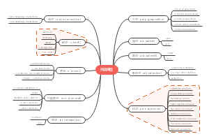
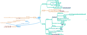
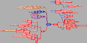
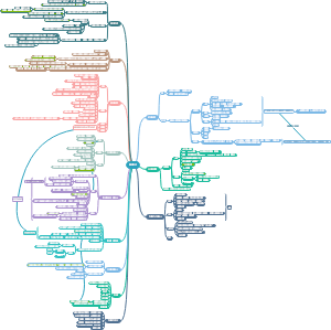
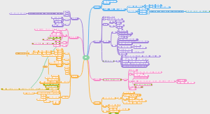
Protein purification
这是一篇关于Protein purification的思维导图,主要内容有The protein we are seperating、1.lon exchange chromatography、2.Gel filtration chromatography、3.Bradford protein concentration method等。
编辑于2022-06-11 15:55:32- Protein p…
- SDSpages
- Bradford
- 相似推荐
- 大纲