导图社区 Membrane proteins
- 11
- 0
- 0
- 举报
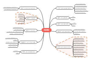
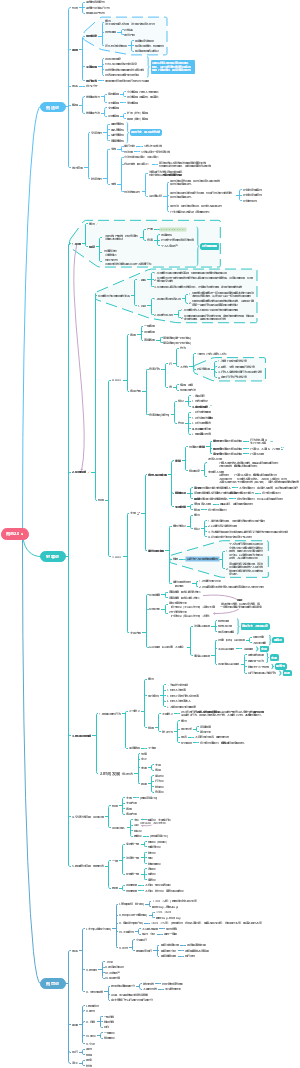
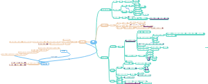
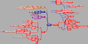
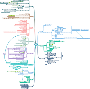
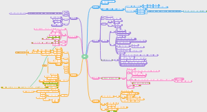
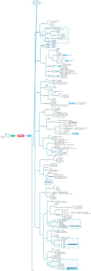
Membrane proteins
这是一篇关于Membrane proteins的思维导图,主要内容有periphral proteins、integral membrane proteins、Synthesis。
编辑于2022-06-11 16:07:52- Membran…
- Synthesis
- cytoskelet…
- 相似推荐
- 大纲
Membrane proteins
periphral proteins
they associates with membrane surface
by electrostatic enteraction with integral membrane proteins.
sugar containing
connected to extracellular mitrix
integral membrane proteins
possible stabalization
specific transmembrane domains embedded them in membrane
transmembrane domains
organizaion
most are free float
some membrane crowed with them, but some sparce
Removal
structure disrupted by excess detergent
modified lipid "ancor" proteins in membrane
nonpolar lipid region
anchor it into membrane
chemical bonding domain
link directly to proteins
adaptation of membrane potential
protein's cytoplasmic side is usually positive charged
protein's extracellular matrix side is usually negative charged
transporters
energy differences
active transporters
Active
against spontaneous process/ let ΔG>0 process happens
most by consume ATP
directly
phosphorylation changes conformation
indirectly
Coupled transport
classificassion
transport one molecule
uniporters
transport two different molecules
in same direction
symporters
in different directions
antiporters
Sodium-potassium pumps
energy cost
1/3 0f energy expended in energy cells
gradient built
higher K+ in; higher Na+ out
so it act as an antipoter
process
3 Na+ bind to cytoplasmic side of protein, and changes protein conformation
This new conformation cleaves ATP
protein phosphorylate it self by cleaving a ATP, and phyosphorylation changes conformation then translocates 3 Na+ across the membrane
This conformation has high K+ affinity, low Na+ affinity
3Na+ dissociate into extracellular fluid
2 K+ bind to protein,and changes it conformation
This conformation hydrolysis phosphate group
hyrolysis phosphate group return protein to its original conformation, translocate 2 K+ and release them to cytoplasma
the origin conformation low K+ affinity, high Na+ affinity
speed
300 Na+ per second
coupled transport protein
strategy
protein capture energy released as one molecule moves down its concentration gradient and use it to move a different molecule against its gradient at the same time.
example
symporter(cotransport)
Intestine epithelial cells intake glucose
transporter use Na+ gradience built by sodium-potassium pump
antiporter(countertransport)
transporter use Na+ gradience transports Ca2+/H+ out
passive transporters
Passive
accelerates spontaneous process by lower activation energy
facilitate diffussion
at first activation energy is high because hydrophobic phospholipids' tail region
lower it by two ways
channel
carrier
1. channel proteins
by the hydrophlic aqueous channel with in the protein
hydrated interior
make it no needs for desolvation,no change on H
selectivity
mostly ions, so we often say ion channels
Ca2+
Na2+
K+
Cl-
some cases more than 1 cation/anion
limiting diameter of channel part
key residues' charge properties face internal of the channel
direction
not defined by structure
so some channel can close/open
3 dominating factors
gate close/open
gated channel only
electrical potential differences
ions only
also called membrane potential/voltage potential
concentration gradience
for all diffussion
gating
gated channel
open/closed by stimulus responses
chemical
ligands
neurotransmiter
electrical
ex. voltage gated ion channels
ungated channel
direction only depends by concentration gradience+potential
ex, normal K+ channels on axon of neuron
kinetics
it should be a Vmax, but it is much higher than carrier's, so linear in most realistic time
reversible, when ΔG=0,a dynamic equilibrium established, both direction's tansport stil accor, but net effect is dynamic equilibrium
2. Carrier proteins
Carrier protien form noncovalent interaction specifically with the molecule they assist
selectivity
ions
small polar molecules
small sugars
aminoacids
noncovalent molecular recognation
most selective
help transport both ions and other solutes
direction
only single direction defined by structure
Kinetics: The rate is determined by
Concentration gradience
normal diffusion is linear relationship
like normal catalyst reaction
but carrier like the kinetics in enzyme
Saturation
carrier has a max turn over number like enzymes
when all carriers are occupied
like Vmax of enzyme
process
1. carrier protein binds the moleclue
2. binding changes carrier's conformation
3. conformation changes bring molecule cross the membrane
4. the molecule dissociates into cytoplasm, and the conformation recovers
irreversible
like enzyme, the reverse reaction is not catalyst/ carrieied
so kinetics should only depends on [S]
So it may act against ΔG
but when ΔG=0, transport stops, with a static equilibrium
it might because the inhibition of conformation change by binding/pressure of the moleclule with in the cytoplasm
doubts?
the inhibition may exist, but would not let it stop at ΔG=0
so what shold you use to against entropy decrease?
binding energy? never it only lower activation enregy(means it must give back to protein)
against second law of thermal dynamics
all natural process must increase universe's entropy
This decrease entropy and no other effects
impossible
so reverse transport must also possible, and also accelerated by same degree
No, an alternative explanation is the reverse transport is always not possible, but the forward transport is blocked by the pressure with in when ΔG=0
。。。 This is another form of equilibrium
not dynamic equilibrium, but pure, static machenic formed equilibium
examples in RBCs
ion carriers
transport Cl- in one direction and HCO3- in the opposite direction
glucose carriers
keep its concentation gradience by converting glucose into charged phosphate glucose in cytoplasm
ΔG's contribution
ions' electrical potential enerygy differences: volts
only accounts when transport ions
also named by membrane potential
entropy differences in concentration
also called concentration gradience
drives diffusion
because of random motion and concentration differences
temperature increase, random motion increase, so diffusion rate increase
classical modle for ΔG=ΔH-TΔS
diffusion stops when concentration sames
don't confuse with solvation, that's most drived by hydrogen bonds formation
loading molecular differences
ions
ion channels
sugars
sugar carriers
water
osmosis
diffussion of water
by free water molecules concentrations differences
which determined by all solutes concentration in a solution
Some definations
hypertonic
The solution with higher concentration
hypotonic
The solution with lower concentration
isotonic
The solutions with same concentrations
osmotic pressure
The force needed to stop osmotic flow
can be meaured as a hydrostatic pressure
strategies to maintain
Extrusion
contract vacuoles in protists
Isosmotic Regulation
high body salts in some marine organisms
cells bathes in an isotonic solution
Turgor
In plants
high salt concentration in central vacules
internal hydrostatic pressure: turgor pressure
aquaporins
water channels
11 kinds found in mammals
some only specific for water
some also allow small hydrophilic molecules
glycerol+Urea
aminoacid
nucleotides
common
both small, hydrophilic molecules(ions/ polar)
contributes to membrane selectivity
receptors
not all receptors are on cell surface
receptor mediated endocytosis
clathrin
The cytoplasmic side pits togeter with embedded receptors act like a molecular mouse trap
mouse trap trigured by proper binding of receptors
specific
fast
tacken up LDL(low density lipoprotein)
which brings cholesteral into the cell
some triggre cellular responces through down stream signaling
identity markers
differenciate cell types
sugar containing
glycolipids
e.x.A,B,O blood types
most
glycoproteins
synthesis
In ER adds sugars of both lipids and proteins
differenciate individuals
MHC proteins
identity proteins distinguish self/nonself by adaptive immune cells
only in vertebrates
highly diverse
physical contacters
junction proteins
usually long-lasting or permanent junctions
tissue specific
determine tissue morphology/architecture
3 types
Tight junctions
they connect plasma membranes of adjacent molecules into a sheet of cells.
ex. the one cell thick digestive tract surface sheet
each cell has
one side out side
transport proteins to absorb nutrients
one side inside to face blood vessels
transport proteins to release nutrients
molecules most can't cross the sheet
tight junctions encircle each cell in the sheet
still have a very slim intracellular space
but no leakage because too tight
hance nutrients must pass through cell
tight junctions partate cell surface, so the different face's transmembrane proteins
Anchoring junction
they covalently attach cytoskeletons between cells or to the extracellular mitrix
most machenical tight
because only connect free-floating membrane is not secure
common in tissues subject to mechanical stress
muscle
skin epithelium
aggregate as a cytoplasmic protein plaque
cytosleletal filaments anchored to plaque
specific proteins types involved
cadherin-mediated links
Desmosomes
connect the cytoskeletons of adjacent cells
Hemidesmosomes
anchor epithelia cells to a basement membrane
Cadherins with in it provide most critical link
most are single pass transmembrane glycoprotein
intracellular part
attach directly to intermediate filament by its short cytoplasmic end
only cadherin can
changes of it is tissue and developmental dependent
ex. migrations of neurons in embryo
attach to actin filament through an attachment protein
more stable
Extracellular domain attaches to another cadherins Through a "hand shake"
Integrin-mediated links
adherens junction
connect actin filaments between cells or to extraceluar mitrix.
mediated by intergins
a large superfamily of cell-surface receptors
they bind to extracellular mitrix proteins
more than 20 different binding domains exist
Communicating junction
these junctions are also channels
channel for small molecules/ions
Chemical/electrical signals can path through it
animals
Gap junctions
composed of connexons
circular complexes of 6 identical transmembrane proteins
diameter 1.5nm
allow path of only small sugars/ammino acids
a channel normally path through plasmamembrane
a lot protruding
dynamic structures
can close/open it self in response to variaty of factors
Ca2+/H+
one function is prevent damage spread between cells
when cells are damaged, leaky plasma membrane increase Ca2+ concentration in cell, and close gap junctions
Ca2+ concentration is normally high out side the cells
when connxons of 2 cells aligned perfectly, they will form gap junctions
connexons spans the plasmamembrane of both cells
plants
plasmodesmata
cytoplasmic connections that form across the touching plasma membrane
contain a central tuble that connects the endoplasmic reticulum of the 2 cells.
occur only in gap/holes between cell walls
where membrane mereges adjacent cell's membrane
built in most living higher plant cells
4 general roles
cell to cell adhesion
both 3 types
cell to matrix adhesion
only anchoring junctions
membrane to cytoskeleton attachment
only anchroing junction
communication & transportation
only communicating junctions
enzymes
Synthesis
Type 1
N-terminus ER lumen
C-terminus cytosol
Type 2
N-terminus cytosol
C-terminus ER lumen
membrane it self
bilayer
5-10nm thick
spontaneouse formation
maximum hydrogen bonding with water
driven by very negative ΔH by forming new hydrogen bonds
and overcome ruduced freedom of motion:negative ΔS
the phosphate head is strongly polar
the fattyacid tail is strongly nonpolar
permeability
It's nonpolar region "repels" polar molecules
not real collumb repels
specifically, barrier because they must temporally get rid of hydrogen boning stabalization to cross the tail region. and at instance of time, it is high in energy.
ΔG it self is negative due to concentration gradience/diffussion
But the process is slow, channel increase the rate by lower this "activation energy"
slow but exist, especially for small polar moleclues like water
big polar molecules are slower than small
Passive transportors facilitate small polar molecules to go through.
facilitate diffussion
lipid components
phospholipid
felxible bilayer mitrix
saturated
less fluidity
no kinks enhance their attchment to form dispertion forces-----pack more tightly
unsaturated
more fluidity
Cholesterol
more in animal cell membrane
rare in plants
increase/decrease fluidity depends on temperature
determine membrane stiffness
LDL brings cholesterol into cell
lipids symmetry
symmetrical
ER
because ER synthesis lipids
asymmetrical
plasma membrane
Golgi apparatus
endosomes
enzymes
enzymes transport lipids across bilayer from one face to the other
factors determine fluidity
temperature
motion make things fluid
dispersion forces
degree of saturation
some bacteria use fatty acid desaturates to aggainst low temperature
deletion of the gene reduces bacteria's cold tolerance
microdomains
plasma membranes are heterogenous
distinct lipids and proteins compose microdomains
e.x. lipid raft
rich in choleseral
cholesteral packs phospholipids more tightly than surrounding membrane
Interior cytoskeleton
structure support
not with in the membrane, underline it but indirectly connect to it
part of cytoskeleton that undernease plasma membrane
contributes to membrane shape
e.x. red blood cell's biconcave shape
specrin scaffold links proteins in plasma membrane with actin filaments in cell's cytoskeleton
anchor some membrane proteins to specific sites
by link to them and control their movement
usually different proteins from strucural support ones