导图社区 Frontier Tech _ Ab RD
- 28
- 0
- 0
- 举报
Frontier Tech _ Ab RD
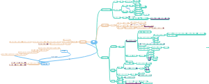
这是一篇关于Ab RD (details mean everythin的思维导图,主要内容包括:Structure,Sources,Quality Control,in vivo assay,Cell based functional assay,Phage based VH/VL screen,Method,Paper,Properity,Protein,Molecule。
编辑于2024-09-18 18:59:25- paper
- Molecule
- 生物医药行业简介
这是一篇关于生物医药行业简介的思维导图,从行业分布、发展趋势、就业方向等角度梳理。主要内容包括:延申,职业,科服,诊断,制药。
- Frontier Tech _ Lifescience _ Ab_Ph.D. Pipeline
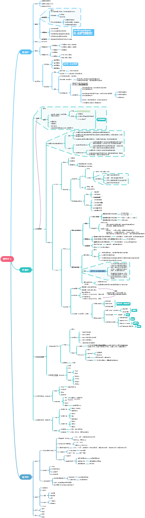
这是一篇关于Ph.D. Pipeline的思维导图,主要内容包括:reported library,Epitope mapping,background,VL library,Fab library,VHH library,Ab label,General Method,reagent,Ab graft,pioneering uses,scFv library。
- Frontier Tech _ Ab RD
这是一篇关于Ab RD (details mean everythin的思维导图,主要内容包括:Structure,Sources,Quality Control,in vivo assay,Cell based functional assay,Phage based VH/VL screen,Method,Paper,Properity,Protein,Molecule。
Frontier Tech _ Ab RD
社区模板帮助中心,点此进入>>
- 生物医药行业简介
这是一篇关于生物医药行业简介的思维导图,从行业分布、发展趋势、就业方向等角度梳理。主要内容包括:延申,职业,科服,诊断,制药。
- Frontier Tech _ Lifescience _ Ab_Ph.D. Pipeline
这是一篇关于Ph.D. Pipeline的思维导图,主要内容包括:reported library,Epitope mapping,background,VL library,Fab library,VHH library,Ab label,General Method,reagent,Ab graft,pioneering uses,scFv library。
- Frontier Tech _ Ab RD
这是一篇关于Ab RD (details mean everythin的思维导图,主要内容包括:Structure,Sources,Quality Control,in vivo assay,Cell based functional assay,Phage based VH/VL screen,Method,Paper,Properity,Protein,Molecule。
- 相似推荐
- 大纲
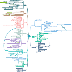
Ab R&D (details mean everything)
1) Correct Result; 2) Shortest Time; 3) Minimum Cost.
Protein
Express
Differences: Expi293: 1) developed by Thermo around 2014; 2) grow faster than CHO; ExpiCHO: 1) gold standard to Ab/Protein production. Some approved drugs(Humira/Avastin/Rituxan) are produced by CHO; 2) CHO has more proper glycosylation modification. Challages (from抗体圈---“从北京到世界:CHO细胞的中国往事”): a, 构建的重组CHO细胞生产效率低,产物浓度低,培养工艺复杂,CHO细胞株稳定性较差; b,某些糖基化表达产物不稳定,不易纯化; c,细胞培养费用昂贵,自动化水平低;
Expi 293F
Expi CHO
hFAP
E.coli
Yeast
Purify (AKTA explorer)
B站视频 官网指导
Size Exclusion Chromatography
Protein A/G resin
Ion Exchange Chromatography (柱材选择)
提高盐浓度,或逆向调pH可洗脱目的蛋白。
Q柱:Quanternary Ammonium, 可结合带负电的蛋白; anion(- 阴离子)
S柱: Sulphonic Acid, 可结合带正电的蛋白; cation(+ 阳离子)
Hydrophobic Column
Feature
Purity(SDS-Page)
Amount(mg/ml-protein/medium)
Thermo stability(4/42℃)
*developability (HPLC) - Retention time
Sepax Zenix-300:(Pmax = 20 MPa, pH 2.0 - 8.5,Rsalt=20mM-2.0M,Rmw=5,000 ~ 1,250,000,Tmax=80℃)
pre-packed Zenix HPLC column 1) Ab prone to aggregate(↓) :: retention time longer :: broader peaks 2) varying the salt content of running buffer :: estimate the nature of interactions between Ab and the column.
TOSOH Butryl-NPR:(Pmax = 20 MPa,Fmax=1.2ml/min,Frec=0.5-1.0ml/min,Falt<0.5ml/min,pH 2.0-12.0,T=10-50℃)
TOSOH TSKgel UP-SW3000:(Pmax =34 MPa, Fmax =0.35ml/min,Frec = 0.1-0.35ml/min,Falt<0.17ml/min,pH2.5-7.5,T=10-30℃)
Modification
一、glycosylation 1, 非岩藻糖的修饰 (afucosylated IgG) 在一些危重的登革热患者体内,IgG抗体的非岩藻糖的修饰 (afucosylated IgG) 丰度是增加的【2】。 抗体依赖性增强效应 (antibody dependent enhancement, ADE) 。简单来说,抗DENV IgG抗体会通过其Fc段与表达Fcγ受体的细胞结合,进而介导病毒进入这些细胞,增加病毒的易感性【1】 2021年6月4日,来自洛克菲勒大学分子遗传学与免疫学实验室的Jeffrey V. Ravetch教授与其合作者们在Science杂志上发表了题为Antibody fucosylation predicts disease severity in secondary dengue infection的研究文章,他们发现非岩藻糖修饰的IgG抗体水平可以用于预测二次感染登革热病毒的危重程度。 IgG1抗体的非岩藻糖修饰水平升高可能与DENV患者的危重程度存在关联性。 IgG1抗体的非岩藻糖修饰水平能更好地反应DENV患者的危重程度。 DENV病毒入侵是特异性地改变了体内IgG1抗体的非岩藻糖修饰水平。 在2020年12月23日,来自荷兰阿姆斯特丹桑昆研究所Gestur Vidarsson教授的研究团队同样是在Science杂志上发表了题为Afucosylated IgG characterizes enveloped viral responses and correlates with COVID-19 severity的研究文章,并与上述研究取得了类似的结论:他们发现COVID-19重症患者体内SARS-CoV-2 IgG抗体的非岩藻糖修饰水平也是升高的,而非岩藻糖修饰的IgG抗体会引起更强的免疫反应,进而触发炎症风暴和急性呼吸窘迫综合征,并最终导致COVID-19重症的产生【3】。 针对这两项研究结果,Science杂志社还邀请了来自新加坡杜克努斯医学院的Ruklanthi de Alwis和Eng Eong Ooi教授在同期Science上发表了题为Antibody sugars are bittersweet的评论文章,对这两个课题组的研究工作都给予了很大的肯定。首先,他们认为Vidarsson教授的研究工作为COVID-19重症患者过度的炎症反应找到了一个可能的解释,即升高的非岩藻糖修饰的IgG抗体有可能触发了这一反应的产生。而Jeffrey V. Ravetch教授的研究工作则拓展了我们对登革热抗体依赖性增强 (ADE) 效应的理解,并找到了一个可以预测感染登革热病毒的危重程度的预后因子。 reference 1. https://science.sciencemag.org/cgi/doi/10.1126/science.abc7303 2. https://science.sciencemag.org/content/371/6532/eabc8378 3. https://science.sciencemag.org/cgi/doi/10.1126/science.abj0435 二、 •2019(Review)-Antibody Structure and Function: The Basis for Engineering Therapeutics.
Graft
Ab graft •2019(Review)-Antibody Structure and Function: The Basis for Engineering Therapeutics.
Concentration
2. BCA assay •Protein concentration was measured by the BCA Protein Assay Kit (Pierce,USA) using bovine serum albumin as the standard.
Structure
Method to determin
single Crystal X-ray diffraction
NMR
Cryo-EM
AlphaFold2
RosettaFold
Method to stablize
site mutation
Ab complex
Ligand complex
Pb (heavy metal)
Properity
Target Binding
Potency of biological modification
Developability
Biological Effects
High expression level
High solubility (at different pH level)
Covalent Integrity
Conformational stability (stable at high Conc./withstand high shear stress)
Colloidal stability (胶体稳定性)
Low poly-specificity (minimal non-specific interaction)
Low immunogenicity
Affinity
Octet(SPR)
Biacore(BLI)
(Label-free)Gator
OpenSPR/Alto(SPR)
Function
Stability
Quality Control
Endo-free(G-_LPS)
remove from liquid
Detergent 可结合 LPS 中的脂质部分,内毒素的脂质部分结合,通过液相分离的方法萃取去除。 对内毒素的去除率高,且不影响有效成份的活性。该法简单高效、价格低廉、适合大规模应用, 在我们日常的蛋白纯化中较为常用。 去内毒素后的样品会有微量 Detergent 的残留。
Triton-114 或 脱氧胆酸钠
SEC
IEX
亲和色谱法
超滤法
0.1%~0.5%活性炭吸附
Physicical
干热:180℃3~ 4h 或 250℃ 30min~ 2h (清除器皿中的)
可溶于水,生产中可用无热原水冲洗以除去热原
可被活性碳吸附
Chemical
0.1M HCl浸泡4h以上
0.1 M NaOH浸泡4hr
30%双氧水浸泡4小时,可完全破坏
3~5%双氧水、重铬酸钾硫酸 清洁液(重铬酸钾∶硫酸∶水常用配比1∶1∶10 或1∶2∶8 )浸泡一般4h以上 (清除器皿中的)
Purity
Amount
Paper
engineered cell line reduce background protein
in vivo assay
Toxicity
mouse
Macaca rhesus
Cell based functional assay
our cel lline
Expresstion
Epi 293F (Expifectamin 293 kit)
Suspension culture
Expi CHO (express hFAP)
Cell Assay
HEK293T _ WT Cell
HEK blue IL2 _ Report Cell (adhension)
CTLL2 _ Proliferation Assay (suspension)
Cell Binding test by Flow Cytometry (eg:HEK293T-hFAP high-E5)
NK-92
how to treat newly arrived cell line? check on Addgene & ATCC.
Cell line construction(for test)
1/ make single resistance cell line; 2/ add another resistance to 1/ cell line;
HEK293F
Method
HPLC (Agilent 1260 infitity Ⅱ)
AKTA
Gator (Probe Life)
Check video
Q-PCR
Multiplate Reader
Snapgene
DNAstar
Isothermal titration calorimetry (ITC)
Phage based VH/VL screen
读书分子细胞、 总结噬菌体展示(文库构建、mapping、细胞系构建、功能验证、表达元件、纯化方案)
Library DNA Design
Library Prep(electroporation)
make competent cells ss320
electroporate library plasmid DNA
megnatic beads-streptavidin + biotin-Ag(lable)
Beads immobilized phage library panning
beads connect target protein-streptavidin
preclean phage by apply onto empeaty beads then bind protein taged beads
wash 3times by PBST/BSA, elute by 200ul glycine(pH2.7),neutralize by 40ul HEPS(pH7.4)
amplify eluted beads&phage respectively by infect XL1-blue(OD600=0.6) 30min, transfer into 30ml 2YT/Amp+/Helper phage
Phage ELISA detect selected VH/VL
Secquencing selected VH/VL
Reconstruct VH/VL to expression in HEK293F
Molecule
Plasmid Construction
successful coloning: 1) pick correst template DNA, ( plasmid, synthesised cDNA, commercial DNA); double check the name and sustance. 2) design proper primer pair: length =18~35mer, 25~35mer better, for long sequence50~70mer; Tm = 55~68, anneal T = Tm-5. Tm=3`- end or after homologous region(≥15bp) or after mutation site; position = mutation/indel near to 5`- end, & 3`- end ≥18mer; anneal = Tm-5, for each pair of primers: ΔT≤4; extention time = depends on length of target fragment, KOD One=5~10s/kb. dpn1 digest (mythelated) template vector befoer in-fusion usually increase positive colone ratio. 3) select correct enzyme here we use "KOD One PCR Master Mix"(Takara) for linearize & amplify, 50ul reaction system; "In-Fusion HD Cloning Kit"(Takare) for construct plasmid, 5ul reacton system, Tm=58~65℃; "Dpn1"(NEB) for digest template vector (contain mythelyted site). 4) proper PCR procedure we activate KOD One at 98℃*5min; In-Fusion: 50℃*15min; 5) correct transformation procedure generally: >> mix in 50ul cells >> on ice ≥20min >> 42℃*1min >> on ice 2min >> 200ul S.O.C * 37℃*250rpm*1hr; Biomiga(倍沃医学) competent cell kit made DH5α can be successful transformed without heat shock. spread 100ul of cultured cells onto LB plate. 6) select proper antibiotics for LB-plate & culture medium Denovo plasmid construct design
Vector:KOD One PCR (template Plasmid DNA ~50 ng / 50 μl)
It`s better to use Dpn1 to digest vector template DNA before run agrose gel. KOD One Polymerase, 50ul reaction system, 2* PCR Master Mix, 引物浓度推荐为0.3 μM(终浓度) Template Plasmid DNA 1 pg~50 ng, recommend 10 ng. Primer Deisgn Len=22~35bp, Tm>63℃; long Frag=25~35bp, Tm>65℃. <三步法循环> 变性 98℃, 10 sec. 退火 (Tm - 5)℃, 5 sec. 延伸 68℃, 1 ~10 sec./ kb 25~45 cycles 延伸时间请根据目的片段长度参考以下方法进行设定: 目的片段长度 建议延伸时间 1 kb以下 1 sec*1 1~10 kb 5 sec / kb*2 10 kb 10sec / kb *1. 有些PCR仪器可能无法设定为1 sec.的延伸时间。扩增量少时,请设定为5 sec。 *2. 如果模板起始量较少或用粗样品进行扩增时,请按10 sec./kb进行设定。 退火(Annealing)温度请按引物的 Tm – 5 ℃进行设定。Tm-5 ℃超过68℃时,请按68℃设定。 DNA变性(Denaturation)推荐98℃, 10 sec.。94℃进行变性时,请设为15 sec.。 Enzyme Failure conditions?
Insertion:KOD One PCR (template Plasmid DNA ~50 ng / 50 μl)
It`s better to use Dpn1 to digest vector template DNA before run agrose gel. KOD One Polymerase, 50ul reaction system, 2* PCR Master Mix, 引物浓度推荐为0.3 μM(终浓度) Template Plasmid DNA 1 pg~50 ng, recommend 10 ng. Primer Deisgn Len=22~35bp, Tm>63℃; long Frag=25~35bp, Tm>65℃. <三步法循环> 变性 98℃, 10 sec. 退火 (Tm - 5)℃, 5 sec. 延伸 68℃, 1 ~10 sec./ kb 25~45 cycles 延伸时间请根据目的片段长度参考以下方法进行设定: 目的片段长度 建议延伸时间 1 kb以下 1 sec*1 1~10 kb 5 sec / kb*2 10 kb 10sec / kb *1. 有些PCR仪器可能无法设定为1 sec.的延伸时间。扩增量少时,请设定为5 sec。 *2. 如果模板起始量较少或用粗样品进行扩增时,请按10 sec./kb进行设定。 退火(Annealing)温度请按引物的 Tm – 5 ℃进行设定。Tm-5 ℃超过68℃时,请按68℃设定。 DNA变性(Denaturation)推荐98℃, 10 sec.。94℃进行变性时,请设为15 sec.。
Gel Extraction (QIAquick® Gel Extraction Kit)
It`s better to use Dpn1 to digest vector template DNA before run agrose gel. QIAquick® Gel Extraction Kit, Quick-Start Protocol, July 2018 vacuum manifold 1) cut gel; 2) 500ul QG, 55℃*10min to dissolve, [ 3 volumes Buffer QG to 1 volume gel (100 mg gel ~100 μl). ] 3) apply onto QIAquick column; 4) vacuum power on; 5) 500ul QG 6) 750 μl Buffer PE optimize: let the column stand 2–5 min after addition of Buffer PE; 7) place column onto 1.5ml tube, 20ul ddH2O, stand for 2min, optimize: 60℃ pre-warmed water may better; 8) centrifuge 13000rpm*1min to elute DNA. optimize: you may reload water again to elute more DNA. Cat No./ID: 28706 QIAquick Gel Extraction Kit (250)
In-Fusion: (5ul system, DNA 50~100 ng)
5ul reaction system: 50℃*15min,4℃hold; Ingredient: Purified PCR fragment: 10–200 ng* Linearized vector: 50–200 ng** * <0.5 kb: 10-50 ng, 0.5 to 10 kb: 50-100 ng, >10 kb: 50-200 ng ** <10 kb: 50-100 ng, >10 kb: 50-200 ng KOD One PCR for V&I, Primer Design: 1) 18–25 bases in length, GC-content between 40–60%; 2) (Tm) between 58–65°C, (Tm) difference ≤ 4℃;(increase length to reach Tm 58–65°C) Note:Tm= the 3’end of the primer, and NOT the entire primer. 3) The last 5 nucleotides at the 3`end of each primer should contain no more than 2 (G) or (C); 4) Avoid: complementarity within each primer to prevent hairpin structures, and between primer pairs to avoid primer dimers; 5) Primer purity: generally use desalted primers; online tools: http://bioinfo.clontech.com/infusion/ Set up the In-Fusion cloning reaction: 5X In-Fusion HD Enzyme Premix 2 µl Linearized Vector x µl* Purified PCR Fragment x µl* dH2O (as needed) x µl Total Volume 10 µl *For reactions with larger volumes of vector and PCR insert (> 7 µl of vector + insert), double the amount of enzyme premix, and add dH20 for a total volume of 20 µl. When designing In-Fusion PCR primers, consider the following: 1. Every In-Fusion primer must have two characteristics: The 5’ end of the primer must contain 15 bases that are homologous to 15 bases at one end of the DNA fragment to which it will be joined (i.e., the vector or another insert). The 3’ end of the primer must contain sequence that is specific to the target gene. 2. The 3’ portion of each primer should: • be gene-specific. • be between 18–25 bases in length, and have a GC-content between 40–60%. • have a melting temperature (Tm) between 58–65°C. The Tm difference between the forward and reverse primers should be ≤ 4°C, or you will not get good amplification. Note: The Tm should be calculated based upon the 3’ (gene-specific) end of the primer, and NOT the entire primer. If the calculated Tm is too low, increase the length of the gene-specific portion of the primer until you reach a Tm of between 58–65°C. • not contain identical runs of nucleotides. The last five nucleotides at the 3’ end of each primer should contain no more than two guanines (G) or cytosines (C). 3. Avoid complementarity within each primer to prevent hairpin structures, and between primer pairs to avoid primer dimers. 4. You can perform a BLAST search to determine if the 3’ portion of each primer is unique and specific (at www.ncbi.nlm.nih.gov/BLAST/). 5. Clontech provides an online tool (at http://bioinfo.clontech.com/infusion/) that simplifies In-Fusion PCR primer design for standard cloning reactions. Simply provide your vector sequence, the restriction enzyme(s) used to linearize the vector (if that is the chosen method for linearization), and the primer sequence required to amplify your region of interest. 6. We generally use desalted oligonucleotide primers in PCR reactions. However, primer quality can de pend on the vendor and varies from lot to lot. If your primer quality is particularly poor (i.e., has many premature termination products), or your primers are longer than 45 nucleotides, they may need to be PAGE purified; however, we usually find this is unnecessary.
Transform:DH5α competent cell (50ul), spred on plate
Transform into 50ul DH5α 1) take out DH5α (50ul/tube, home-made with BW-SCCP kit) from -80℃ stock, thaw it on ice around 2min; (not thaw completely) 2) add 5ul(no more than 1/10 of cell volume) in-fusion product into 50ul DH5α tube, tap the tube very gently around 5 times, stay on ice around 25min; 3) heat shock at 42℃ for 1min; for some competent cells, this step is not necessary. [we can ommit it here with BW-SCCP-prepared DH5α cells] 4) immediately put the tube on ice for 2min just after heat shock; [any move or shake may decrease transformation efficiency] 5) add 200ul S.O.C medium into the tube, incubate at37℃*250rpm*more than 30min; 6) pipete 100ul of recovered cells onto 37℃ pre-warmed LB plate with proper antibiotic resistance. (Generally, we use LB-amp plate.) 7) incubate plate at 37℃ over night. (around 16hr) 8) Conone-seq: pick colonies & Incubate in 200ul 2YT(amp) ≈3hr, seq to TP-seq. (If correct do mid prep & midi-seq.) 9) Midi culture: pipete 100ul of single colony culture into 30ml 2YT(amp) incubate 37℃*250rpm for 16hr; (100ul subculture may got 5000ng/ul eluted by 50ul ddH2O, and 50ul may got 1500ng/ul) 10) Midi prep: Plasmid Midi Prep (QIAGEN Plasmid Plus Midi Kit); 11) If midi-seq result is correct deliver a tube of plasmid with around 1000ng/ul to Yang for transfection/expression & purification/funcitional assay.
Site-Directed Mutageneiss
Site mutation [Indel / substitution] Primer Design matters a lot! Use correct template DNA . Design & use correct primer pairs. 1) the mutation site should be centrally located on both primers of each pair; 2) primers purified by desalting usually work well, desolve in DNA- & RNA-free water; 3) Mutagenic primers must have unique binding sites; 4) complementary sequences between primers must be avoided. 5) Primers that have a similar Tm and a G or a C at their 5`- end work better; 6) Each multi-site mutagenesis primer must contain 15 nucleotides of non-mutagenic sequences flanking the outer-most modification(s) (i.e., mutations on the 5’ and 3’ position of the mutation site). 7) If there are ≤50 nucleotides between two mutation sites, we recommend using a single mutagenic primer pair to introduce them into the target plasmid; Do with correct PCR procedure. Dpn1 digest template (mythelyted) DNA. transform & incubate proper time to pick & seq-check single colonies. 1 or 2 sites mutation don`t need to deactivate Dpn1 after digest template vector by it.
VL/VH insert stop codon
library design
ssDNA Prep with CJ236 bacteria
Kunkel Site-directed Mutaion
we have done: 1) D047/49/64 + "TAATAA", (Replace by stop codon) 2) V868/69/70 + "TAATAA". (insert stop codon) Luke designed Kunkel Mutation Primers by introduce"NKK". Command Kunkel 1, Site-Directed Mutagenesis - Kunkel Method (with chart) 2, 2012-Improvements to the Kunkel mutagenesis protocol for constructing primary and secondary phage-display libraries; 3, 2019-Improving the mutagenesis efficiency of the Kunkel method by codon; 4, CV of Thomas Kunkel; 5, 1985-Rapid and efficient site-specific mutagenesis without phenotypic selection. Compare Kunkel & PFunkel mutation method 2012-PFunkel: Efficient, Expansive, User-Defined Mutagenesis (Plos One) Why site-mutation method hasn`t been break since 1985,Kunkel?
prepare competent SS320 cells for electroporation
Refer to "Electroporation Protocol" Aim To make a library of pahge contain huge amount of variable region for screen. Brief Procedure calculate amount of cells need; streak ss320 onto LB-tet plate, culture single colony to reach log phase (OD600=0.5~0.8), harvest cells in all cold condition, got small volume and high concentration electroconpetent cells ; electroporation (purify kunkel product before electroporation) ; detect titer of electroporated transformants (calculate transform efficiency); phage production & isolation ; NGS to detect libary diveristy ; optimize efficiency & diversity based on cells/DNA amount; Key Points calculate amount of competent ss320 needed to electroporate according to how much plasmid/kunkel products wants to transfect; preparations : 1) Kunkel mutation production, (mutation primers PCR, phosphoralation, circlize ligation)to got library, purify kunkel production before electroporation by *** kit; (spread onto LB-amp plate to confirm kunkel product is good) 2) Ice, (everything touch preconpetent cells need to be precool), electroporation cuvette (1mm or 2mm ), precooled ddH2O, 【3 times wash with 1mM HEPES, 4th time wash with pure ddH2O】 Superbroth (Yeast, Tryptone, Glycerol, K2H-KH2 solution) to prepare ss320, S.O.C. (Yeast, bacto tryptone, NaCl, KCl, MgCl2, MgSO4, Glucose) to recover electroporated ss320, 2YT (Yest, Tryptone, NaCl, pH7.0) to subculture electroporated ss320 , 3) prestreaked ss320 LB-tet plate, (can keep 2 weeks at 4℃ for use), pick single colony to culture in 2YT to log phase(OD600=0.5~0.8), electroporation condition: 1) 2mm cuvette: 2300KV*5ms, 【DNA=560ng/ul, 4ug≈7ul; 400ul/tube ss320; NC, PC in parallel】; 2) 1mm cuvette: 1800KV*5ms, 【≈500ng DNA/80ul conpetent cells,】 Note: the amout of library DNA and cells would be vary according to optimized procedure. 3) mix library with prepared ss320 cells, keep on ice for 5min,electroporate; 4) immediately add 1ml S.O.C to cuvette, then transfer into 14ml tubes,wash cuvette with another 4ml S.O.C. and also transfer to the 14ml tubes, incubate 37℃*1hr*200rpm; 5) subculture each tubes electroporated ss320 into 250ml flask contain 30ml 2YT(amp/kan), incubate 37℃r*250rpm over night. 6) do titering to evaluate number of transformants. subculture electroporated ss320 & extract phage, sequencing to detect electroporation efficiency & library diversity. V: Viability, TE: Transfection Efficiency. http://www.nepagene.jp/e_products_nepagene_0002.html
pick 1~3 colonies from pre-streaked plate into 2ml 2YT to OD600=0.6(≈3hr)
M13KO7 helper phage production
Phmid plasmid construction
Library Screen (3~5rounds)
1st Round Screen: all the eluted phage are used to infect host cell(XL1-Blue): then, 1) Infection: 100ul Glycine eluted phage + 20ul 1M HEPES(1/5 V) + 500ul log phase XL1-blue cells, incubate at least 20min at RT; 2) Titering: 10 times serial dilution by ddH2O, (mind the dilution times calculation). plate these on LB(amp) plate, 37℃**over night; [for 6*6 square plate, we load 10ul/square, e3, e4, e5, e6, e7, e8 ] 3) Sub-culture: 14ml tubes = 2.5ml 2YT + amp + M13K07 helper phage + eluted phage; 37℃*250rpm*over night; Other Round Screen: after elution & neutralization, got 120ul of eluted phage library. >>> take 50ul eluted phage + 500ul mid log phase XL1-blue (host) cell, [done 2 times dilution] incubate at least 20min at RT; >>> keep the rest of eluted phage in EP tube and label for use; >>> take 10ul of infected cells to serial dilution (eg: 990ul ddH2O) [done 50 times dilution] >>> sub-culture phage for next round of screen,
Dilute phage lib & Ag
mind the calculation of dilution. Phage library dilute to A268=0.2 with PBST/BSA (0.2%tween20, 0.5%bsa). Ag = EPCAM (#1,3,9,11,12) Trop2 (#15) dilute to 10nM with PBS.
Ag-Biotin-Streptavidin-Beads
Screen in 96-well plate with 09092019SelectionProtocol, load 100ul/well. Ag dilute with PBS to 10nM, Beads 2ul/well; wash twice with PBS before use. Biotin 10mM, 100ul/well. "09092019SelectionProtocol" (61min) A == Ag( variable [ ] ) + beads (2ul) B == Biotin Solution (10uM) C == Phage (A268=0.2) D == PBST/BSA (0.2% v/v tween20, 0.5 m/v bsa) E == PBST/BSA F == PBST/BSA G == PBST/BSA H == Glycine (100mM,ph2.7) ——————this protocol run by KingFisher.
Screen Protocol
Screen in 96-well plate with 09092019SelectionProtocol, load 100ul/well. Ag dilute with PBS to 10nM, Beads 2ul/well; wash twice with PBS before use. Biotin 10mM, 100ul/well. "09092019SelectionProtocol" (61min) A == Ag( variable [ ] ) + beads (2ul) B == Biotin Solution (10uM) C == Phage (A268=0.2) D == PBST/BSA (0.2% v/v tween20, 0.5% m/v bsa) E == PBST/BSA F == PBST/BSA G == PBST/BSA H == Glycine (100mM,ph2.7) ——————this protocol run by KingFisher. + 20ul 1M HEPES after selection to well-H to neutralize sample.
Competition Protocol
Screen in 96-well plate with 09092019SelectionProtocol, load 100ul/well. Ag dilute with PBS to 10nM, Beads 2ul/well; wash twice with PBS before use. Biotin 10mM, 100ul/well. "15min Off Rate Protocol" (75min) A == Ag( variable [ ] ) + beads (2ul) B == Biotin Solution (10uM) C == Phage (A268=0.2) D == PBST/BSA (0.2% v/v tween20, 0.5 m/v bsa) E == Competitior (1uM) F == PBST/BSA G == PBST/BSA H == Glycine (100mM,ph2.7) ——————this protocol run by KingFisher. + 20ul 1M HEPES after selection to well-H to neutralize sample.
Materials
Bacteria
XL1-blue (Amplify Phage)
CJ236 (Prep ssDNA)
M41061
ss320 (Electropor. Competent)
DH5α (Chem. Competent)
Top10 (Chem. Competent)
Phage
Phagmid Helper phage: M13K07
M13KO7 helper phage
Phagemid
Kits
Sources
Single B cell
memory B cell secreted Ab
PBMC
Yeast display
Cell display
Tumor cell
cancer cell secreted Ab
Record Kits/Method/Key Points/
1st Month Report
Ab
Protein A Purify
Monofinity A Resin (GenScript)
PBS
Wash Buffer
0.5M Nacl PBS wash at least 10 times of resin volume
Elution Buffer
0.5M Glycine (ph3.0), 10ml * 2 times preload Neutralize buffer in collection tube: 1M Tris (pH8.0), 1/10 volume of Elution volume. (endo-free) What`s the mechanism of Glycine to elute Ab/protein? the same as elute phage while screen library.
Resin
Volume to use Regeneration: at least 2 column volumes of 0.1 or 0.5 M NaOH with a contact time of 10–15 minutes, use 0.1 M NaOH every cycle and 0.5 M NaOH every 10 cycles PBS wash
SDS-Page
Reduce/Non-Reduce
Molecular Weight Analysis
Detect Conc. & Dialysis
Developability (Agilent 1260 Infinity)
Injection Volume = 10ul; Control Panel: Pressure Alarm; Fow Rate; Stop Time/Volume; Sampling Sequence -- a, Acq Method b, Sample name c, Data file (result name form) d, Result Path e, Result Name Shut down Program; Baseline stable at 0 mAu; Correct match: Buffer & Inject ; Sample & Sequence Position; Anayssis: Retention Time; Q: 用什么柱子分析了什么蛋白,上样量是多少,出峰位置是多少?阳性对照是什么?实验结果是什么?结论是什么?
Sepax Tech. Zenix SEC-300
Retention Time Longer >> Ab may stick onto column(stationary phase) material; Retention Time shorter >> Aggregation/Precipitation Buffer:
TOSH-TSKgel UP-SW3000
TOSH-TSKgel Butyl-NPR HIC
Hydrophobic(疏水) Interaction Chromatography Retention Timer Longer >> hydrophobic residues Increase/ Exposure; Retention Timer Longer >> Hydrophilic residues Increase/ Exposure; Buffer:
Thermo Stability
4℃/42℃ -- 2 month weekly detect Melting Curve
Principle Operation Data Analysis
AKTA
Practice more. Bach purification
Q/S column
结合什么蛋白,就叫什么柱。 S = strong cation(阳) exchanger, sulphonic acid functional groups; 蛋白(+),-磺酸基团 Q = strong anion(阴) exchanger, quaternary ammonium functional groups. 蛋白 (-),-季铵基团 # 阳离子柱(S):结合带正电的蛋白,洗脱带负电的杂蛋白; 阴离子柱(Q):结合带负电的蛋白,洗脱带正电的杂蛋白。 # eg: A protein with pI 9 is positively charged at pH 7 and will bind to the cation (S) columns. Increasing the salt concentration or raising the buffer pH above 9 will elute the bound protein. (Protein will negetively charged at pH>9,so that protein can`t bind to cation column for they both negetively charged) A protein with pI 4 is negatively charged at pH 7 and will bind to the anion (Q) columns. Increasing the salt concentration or lowering the buffer pH below 4 will elute the bound protein.
Molecular
Cloning
KOD One PCR for Insertion/Vector fragments, 50ul system, 10s/kb; >> Gel Extraction
In-Fusion, 5ul system; >> Transform into DH5α
Colony Culture & Seq >> Midi Culture & Seq
Library Prep
ssDNA Prep (CJ236)
Kunkel site-directed Mutation
XL1-blue: 100ml to OD600=0.6
XL1-blue cells are prepared for phage infection, during titering library phage. Log phase ≈ OD6000.5~1.0 .
electroporation
special materials
Ice
cold: ddH2O(with/without HEPES), cuvette, tips, centrifuge bottle,
ddH2O wash 3 times with HEPES, 4th ddH2O wash without HEPES.
warm: SOC
Medium: Superbroth, 2YT, SOC,
Superbroth: for ss320 SOC: recover after electroporation 2YT: grow recovered cells
Prep ss320 strain
Resistance: ss320 --- Tet Helper Phage --- Kan Library Plasmid DNA --- amp
Electroporate: Lib-DNA & ss320 competent
0.2cm cuvette
no salt, 300~400ul cells: 4ug DNA, 2500 kV, 5ms + SOC immediately, recover 1hr Titering
0.1cm cuvette
100ul cells: 80ng DNA 1800kV, 5ms
recover 1hr
+ 1ml 37℃ pre-warmed SOC, 37℃*220rpm*1hr
Amplify(什么时候为什么扩增什么)
subculture the transformated ss320 at least 2:1 to starting volume == eg: 1L ss320 in Superbroth, subculture into at least 2L of 2YT(correct antibiotics) for phage growth. mesure OD600 ~0.7, + M13K07 to a final Conc.=10e10 pfu/ml; incubate ~30min at 150rpm*37℃, 250rpm**37℃, over night.
Titering 实际在多少稀释倍数下数的多少个plaque?
PC: into 10ml SOC, NC: 400ul ss320 dilute 100 times, spread onto plate; Sample: in 100ml SOC, to dilution dilution times: e-5, e-7, e-9, e-10,
PC: electroporate plasmid DNA, same amount
PC: electroporate with plasmid DNA, recover with 10ml SOC,
NC: ss320 without electroporate, ?same amount(正确用量多少);NC的意义/作用是什么?
Dilution: 10e-5, 10e-7, 10e-9, 10e-10,
Plate: LB(amp), 6*6 squar plate
M13K07 helper Phage(Learn how to prepare/use it?---protocol)
1st: pellet cells 6500rpm*1hr 2nd: 6500rpm*30min resuspend by 20~40ml PBS, JA-10 rotor: 10000g*15min, precipitate phage: +1/5 volume of PEG/NaCl, mix, keep on ice 30min, pellet phage: 10000g*20min.
Library Screen
Principle Operation Analysis Trouble Shooting Practice more than 10 times What`s the mechanism of Glycine elute phage while screen library? the same as to elute Ab/protein while purification.
KingFisher
Round 2 (2020.10.21)
Material
Medium (Ingredients of Superbroth, 2YT?)
Superbroth SOC 2YT LB-Agar
Superbroth
2 YT
S.O.C
LB (Liquid/Plate)
Antibiotics (which strain has what resistance)
Amp Kan Chl Tet
Strain
a. CJ236: LB-Agar (Chloramphenicol Resistance) b. XL1: LB-Agar (Tetracycline Resistance) c. ER2738: LB-Agar (Tetracycline Resistance) d. MC1061: LB-Agar (Streptomycin Resistance) e. SS320: LB-Agar (Streptomycin + Tetracycline Resistance) f. TG1: Minimal Media-Agar (no antibiotic) g. DH5: LB-Agar (no antibiotic)
Buffer
PBST/BSA PEG/NaCl PBS ddH2O
Need to learn
Gator
Principle Operation Analysis Trouble Shooting Practice more than 10 times Coach 3 other me
Multiplate Reader
Principle Operation Analysis Trouble Shooting Practice more than 10 times
Flow Cytometry
Strengthen
Ab
Thermodynamics
Design Operation Analysis
AKTA batch purification
Injection valve shift; Control Panel: a, buffer chanel selection; b, column selection;
Molecular
Connect ssDNA prep and electroporation
Kuncel mutation
Screen
Ab Label & Test (eg:biotin……)
Tian mentioned NHS method to label. N-hydroxysuccinimde (NHS) N-羟基琥珀酰亚胺酯
Combine Protein onto Magnetic Beads
浮动主题
when it comes to "Learn" Principle Operation Analysis Trouble Shooting Practice more than 10 times Coach 3 other me